Introduction and Context
Left main coronary artery (LM) disease remains one of the most consequential lesion subsets encountered in contemporary interventional cardiology. The left main supplies a large myocardial territory; disease here carries heightened procedural risk and a greater likelihood of late adverse events after percutaneous coronary intervention (PCI). Over the last decade, growing evidence has shown that intracoronary imaging—in particular intravascular ultrasound (IVUS) and optical coherence tomography (OCT)—can materially improve procedural decision-making and outcomes in complex PCI. Against this backdrop, a global expert panel coordinated by the European Association of Percutaneous Cardiovascular Interventions (EAPCI) in collaboration with the European Bifurcation Club (EBC) published a clinical consensus statement (Johnson et al., Eur Heart J. 2026) focused specifically on intracoronary imaging for left main bifurcation PCI.
Why this consensus now? Several clinical gaps and developments prompted the document:
– New randomized and registry data strengthen the case that imaging-guided PCI reduces restenosis and adverse events in complex lesions, including left main disease.
– Uptake of imaging is variable internationally; a focused consensus aimed to translate evidence into clear, actionable recommendations for planning, guidance and optimization of LM PCI.
– The left main bifurcation presents unique anatomical and technical challenges (ostial disease, large vessel size, frequent involvement of the LAD/LCx bifurcation) that benefit from imaging-specific algorithms and thresholds.
The rest of this article summarizes the core recommendations, highlights what is new compared with prior guidelines, presents topic-by-topic guidance, and offers practical commentary for clinicians and health systems.
New Guideline Highlights
Major themes and takeaways from the 2026 EAPCI–EBC consensus statement
– Strong endorsement of intracoronary imaging (IVUS or OCT) as the standard of care for left main bifurcation PCI: imaging is recommended for lesion assessment, device sizing, stent optimization and final result evaluation.
– Imaging should be integrated into all phases of care: procedural planning (anatomic characterization and strategy selection), procedural guidance (landing zone and sizing), and optimization (post‑dilatation, assessment of stent expansion, malapposition, edge dissection and residual disease).
– Practical algorithms and segment-specific imaging targets are provided for decision-making at the distal left main bifurcation and for ostial LM lesions.
– Recognition of the different strengths of IVUS and OCT: IVUS favored where greater penetration and sizing in large vessels are required; OCT useful for detailed appraisal of stent strut apposition and edge dissections where blood clearance can be performed safely.
– Emphasis on training, quality assurance and device access — the committee argues that patient benefit will only follow if operators and centres embrace imaging-based workflows and competency standards.
Key condensed recommendations (high level)
– Class I recommendation: Use intracoronary imaging (IVUS or OCT) to guide LM PCI planning and optimization in both acute and chronic coronary syndromes where PCI is pursued.
– Class I recommendation: Use imaging to confirm adequate stent expansion and to identify significant residual issues that require correction (malapposition, underexpansion, edge dissections).
– Practical preference: IVUS is often preferred for large-caliber left main vessels; OCT is complementary when high-resolution assessment of struts or dissections is required.
Updated Recommendations and Key Changes
How the 2026 consensus differs from previous guidance
– Stronger, more prescriptive imaging endorsement: Earlier revascularization guidelines and position papers increasingly acknowledged benefits of imaging for complex PCI, but the 2026 statement moves from recommendation to operational guidance, with detailed stepwise algorithms for LM lesion types.
– Focused left main bifurcation content: Prior general PCI imaging guidance treated coronary territories collectively. The EAPCI–EBC consensus supplies left main–specific appraisal, including bifurcation strategy integration (provisional single-stent vs planned two-stent approaches) and imaging thresholds tailored to LM anatomy.
– Practical implementation emphasis: the statement addresses real-world barriers—training, procedural time, reimbursement—and makes recommendations for clinician competency and institutional adoption pathways.
Table (summary of changes versus earlier guidance)
– Previous guidance (e.g., ESC/EACTS myocardial revascularization guidelines): supported imaging for complex PCI with Class IIa/IIb recommendations in many contexts.
– 2026 EAPCI–EBC consensus: elevates intracoronary imaging to a core component of LM PCI workflows (operational Class I-style endorsement for planning and optimization), and provides segment-specific algorithms and MSA-focused optimization targets.
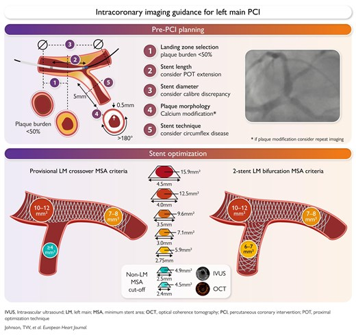
(Note: for the full list of numeric MSA thresholds, operator algorithms and boxed stepwise protocols consult the full Johnson et al., 2026 consensus statement.)
Topic-by-Topic Recommendations
1) Indications and patient selection
– Who should get intracoronary imaging for LM PCI?
– All patients undergoing PCI for significant left main disease (ostial, body, or distal/bifurcation) — imaging is recommended to support procedural planning and to optimize stent result.
– Imaging is particularly crucial in cases with angiographic uncertainty (intermediate stenosis), heavy calcification, large vessel diameter, or bifurcation involvement.
– In acute coronary syndromes involving the left main, imaging should be used when feasible after initial stabilization to confirm lesion morphology and guide stenting decisions.
2) Imaging modality choice: IVUS versus OCT
– IVUS strengths and recommended uses
– Better penetration in large-caliber vessels such as the left main → preferred for vessel sizing and plaque burden assessment.
– Less affected by residual blood; advantageous in ostial LM lesions and when vessel size exceeds OCT field.
– OCT strengths and recommended uses
– Much higher axial resolution → superior for detecting small dissections, malapposition at the strut level, and tissue prolapse.
– Requires blood clearance and careful contrast management; may be less practical in large LM diameters or in hemodynamically unstable patients.
– Practical approach: use IVUS as the first-line imaging tool for most LM procedures; use OCT as an adjunct when high-resolution assessment of stent apposition or subtle dissections is needed.
3) Planning and procedural guidance
– Use imaging pre-intervention to:
– Confirm significance of angiographically intermediate LM lesions, avoiding unnecessary interventions.
– Measure reference vessel diameter and lesion length to choose stent diameter and length.
– Identify heavy calcification, need for atherectomy, or other plaque modification strategies.
– Bifurcation strategy selection
– Imaging helps define the true bifurcation anatomy (carina position, plaque distribution) and therefore supports choice of provisional single stent versus two‑stent techniques.
4) Optimization and minimal stent area (MSA) targets
– Core principle: stent underexpansion is a dominant mechanism of restenosis and stent thrombosis. The consensus provides MSA-based targets (segment‑specific) to guide post‑dilatation and optimization.
– Practical guidance (summary):
– After stent deployment, perform imaging to measure MSA and inspect for malapposition and edge dissection.
– If MSA is below recommended segment thresholds or if significant malapposition/flow‑limiting dissection is present, perform further high-pressure post‑dilatation and reassess.
– Operators should use balloon sizes and pressures guided by imaging-derived reference dimensions rather than angiographic estimation alone.
(For full numeric MSA thresholds by LM segment and clinical scenarios, see the consensus text; the authors provide validated segment-specific targets and escalation pathways.)
5) Ostial LM disease
– Ostial lesions are particularly challenging due to the need for precise proximal landing against the aortic wall.
– Imaging (especially IVUS) is recommended to confirm true ostial disease, to guide accurate proximal stent positioning, and to assess for geographic miss.
6) Post‑procedure surveillance and follow-up
– Document intravascular imaging findings in procedural records to guide follow-up; imaging-defined underexpansion or residual disease may prompt closer clinical surveillance.
– Use imaging data to inform secondary prevention intensity (e.g., dual antiplatelet therapy duration after complex stenting may consider imaging findings such as malapposition or edge dissection).
7) Special populations
– Acute coronary syndromes: imaging is recommended when hemodynamics permit; OCT may be limited in very unstable patients because of contrast needs.
– Renal impairment: minimize contrast load; IVUS preferred because it does not require additional contrast for imaging.
– Very large vessels: IVUS preferred because OCT axial field and penetration may be insufficient.
Recommendation Grades and Practical Quick Reference
– Core recommendations (condensed):
– Use IVUS or OCT to plan, guide and optimize LM PCI — strong endorsement from the consensus group (operationally equivalent to a Class I recommendation for routine use in LM PCI).
– IVUS favored for vessel sizing and ostial/large-vessel assessment; OCT complementary for high-resolution evaluation of strut apposition and dissections.
– Obtain post-deployment imaging and correct any identified underexpansion or clinically important complications.
– Implementation checklist for operators:
1. Pre-PCI imaging to confirm lesion significance and to measure reference diameters.
2. Use imaging to choose stent diameter and length; plan bifurcation technique.
3. After stent deployment, perform imaging to measure MSA and check for malapposition or edge issues.
4. Optimize with targeted post-dilatation and confirm with repeat imaging.
5. Record findings and optimization steps in procedure notes for follow-up planning.
Expert Commentary and Insights
What did the committee emphasize beyond the core recommendations?
– The consensus authors stressed that the patient benefit shown in multiple datasets will only be realized if the interventional community adopts imaging-guided workflows broadly. Training and competence matter: reading intracoronary images and translating them into corrective steps requires experience.
– Cost and resource implications were discussed explicitly. Imaging adds equipment cost and procedural time, but proponents argue that by reducing restenosis, target lesion revascularization and stent thrombosis, imaging can be cost‑effective in large or complex coronary interventions, particularly left main disease.
– Controversies highlighted by the panel:
– The evidence base includes randomized trials and robust registries for general complex PCI, but fewer randomized trials are focused solely on left main PCI—hence the consensus approach synthesizes best available data and expert experience.
– Choice between IVUS and OCT remains context-dependent; the group avoided an absolute preference but provided practical scenarios favoring each modality.
– Research priorities identified by the panel include randomized trials comparing imaging-guided versus angiography-guided left main PCI with contemporary stents and adjunctive therapies, and trials addressing imaging-driven thresholds for escalation of optimization maneuvers.
Practical Implications for Daily Practice
– Operator-level changes
– Expect to use imaging as part of routine LM PCI workflows: pre-PCI assessment, sizing, and post-PCI optimization should incorporate IVUS/OCT.
– Allocate time and training to achieve competence in image interpretation and optimization techniques.
– Institutional/system implications
– Healthcare systems and catheterization laboratories should consider investment in imaging consoles, disposables, and staff training, and adapt scheduling and billing pathways to reflect imaging‑enhanced procedures.
– Quality programs should track imaging use and outcomes: % of LM PCI cases with imaging, rates of post‑optimization MSA achievement, and longer-term clinical endpoints.
– Patient communication
– Explain to patients that intracoronary imaging provides detailed inside-the-artery information that informs safer, more precise stent placement and reduces the risk of recurrent problems.
Practical vignette
Michael, a 68-year-old man with exertional angina and severe three-vessel disease, is found to have a 60–70% angiographic stenosis of the distal left main involving the bifurcation. Before committing to PCI, the operator performs IVUS to confirm lesion severity, measure the reference diameters and define plaque distribution. The IVUS shows a large-caliber LM with eccentric plaque extending into the proximal LAD; the operator selects a provisional single‑stent strategy sized to IVUS-derived reference diameters, deploys the stent, and then uses IVUS to identify focal underexpansion at the distal stent edge. High-pressure post‑dilatation is performed and repeat IVUS confirms the MSA target has been met. Michael recovers without complication and avoids early repeat revascularization.
Conclusions
The 2026 EAPCI–EBC clinical consensus statement makes a clear, practice-oriented case that intracoronary imaging (IVUS or OCT) should be an integral component of left main bifurcation PCI. The document elevates imaging from a recommended adjunct to a fundamental tool for planning, guiding and optimizing interventions in this high‑risk coronary territory. Successful translation into improved patient outcomes will depend on operator training, institutional support, and further prospective research to refine imaging-derived targets and pathways.
References
1) Johnson TW, Gonzalo N, de la Torre Hernandez JM, Amabile N, O’Kane P, Alasnag M, van Geuns RJ, Holm NR, Brugaletta S, Raber L, Albiero R, Ferenc M, Pan M, Paradies V, Ali ZA, Jia H, Lemos P, Mathew R, Zhang JJ, Hahn JY, Chiatzizisis YS, Burzotta F, Banning AP, Guagliumi G, Hildick-Smith D, Chieffo A, Akasaka T, Mintz G, Louvard Y, Flensted Lassen J, Stankovic G. Intracoronary imaging for left main percutaneous coronary intervention: a clinical consensus statement of the European Association of Percutaneous Cardiovascular Interventions (EAPCI) of the ESC and the European Bifurcation Club (EBC). European Heart Journal. 2026 Oct 5;47(37):5247-5265. PMID: 42135060. URL: https://pubmed.ncbi.nlm.nih.gov/42135060/
2) Neumann FJ, Sousa-Uva M, Ahlsson A, et al. 2018 ESC/EACTS Guidelines on myocardial revascularization. European Heart Journal. 2019;40(2):87–165. (European Society of Cardiology / European Association for Cardio-Thoracic Surgery guideline that previously addressed imaging in complex PCI.)
(Note: The full EAPCI–EBC consensus provides detailed numeric MSA thresholds, segment-specific algorithms and boxed procedural checklists. Clinicians performing left main PCI should consult the complete consensus document for the precise imaging-derived targets and stepwise optimization protocols.)