
Highlights
- Elevated baseline body mass index (BMI), especially in overweight and obese categories, associates with improved overall survival in head and neck cancer patients undergoing radiotherapy.
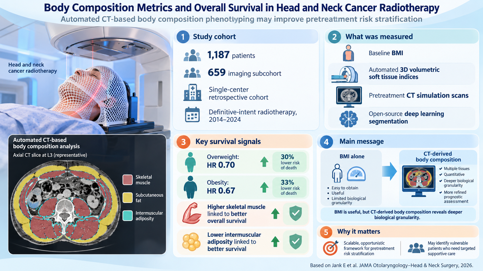
- Automated three-dimensional CT-derived body composition phenotyping reveals that higher skeletal muscle mass contributes to better survival outcomes, while elevated intermuscular adiposity correlates with increased mortality risk.
- AI-assisted cervical muscle assessments (e.g., FUNC-RISK score) offer promising prognostic stratification tools derived from routine radiotherapy planning CT scans.
- Combining classical clinical nutritional markers (e.g., serum albumin, handgrip strength, prognostic nutritional index) with imaging-based body composition improves risk prediction and informs targeted supportive interventions.
Background
Head and neck cancers (HNC) represent a diverse group of malignancies with considerable morbidity and mortality despite advances in multimodal therapies, including definitive radiotherapy and chemoradiotherapy. Accurate prognostication is critical to optimize therapeutic decisions and supportive care to mitigate treatment-related toxicities and improve survival outcomes. However, heterogeneity in patient frailty, nutritional status, and body composition complicates uniform risk assessment. Traditional metrics such as body mass index (BMI) offer an accessible but limited snapshot of nutrition and body composition. Emerging evidence implicates sarcopenia (loss of skeletal muscle mass and strength) and adiposity patterns as more nuanced biological markers that influence treatment tolerance and oncologic outcomes. Recent advances in automated imaging analysis, including volumetric three-dimensional phenotyping from computed tomography (CT), enable objective quantification of body composition beyond conventional anthropometric measures. This review synthesizes recent high-quality evidence elucidating the independent prognostic roles of baseline BMI and CT-derived body composition metrics—including skeletal muscle, subcutaneous, and intermuscular adipose tissue—in patients with head and neck cancer undergoing radiotherapy. It further explores complementary functional assessments and biochemical markers, integrates methodological developments involving AI-assisted phenotyping, and discusses translational implications for personalized oncologic care.Key Content
Baseline BMI and Survival Outcomes in Head and Neck Cancer Radiotherapy
A retrospective cohort study by Jank et al. (2026) analyzed 1187 patients treated with definitive intent radiotherapy for head and neck cancer, revealing that overweight (HR 0.70; 95% CI, 0.56-0.89) and obesity (HR 0.67; 95% CI, 0.50-0.89) categories independently predicted improved overall survival compared with normal weight. This protective association aligns with prior reports indicating an ‘obesity paradox’ in oncologic contexts, potentially reflecting reserves to withstand treatment toxicity and disease cachexia. Nutritional biomarkers such as serum albumin and prognostic nutritional index (PNI), low levels of which predict treatment interruption and survival decrements, further corroborate BMI’s clinical significance (Martins et al., 2026; Karakus et al., 2025). However, BMI expansion predominantly reflected increased subcutaneous adiposity rather than skeletal muscle gain, underscoring the limited granularity of BMI alone for prognostic modeling.Advanced CT-Derived Body Composition Phenotyping
Automated 3D volumetric assessments using open-source deep learning segmentation pipelines from pretreatment CT simulation scans permit quantification of skeletal muscle, subcutaneous, and intermuscular adipose compartments. Within an imaging subcohort (n=659) of the Jank et al. study, elevated skeletal muscle volume significantly improved overall survival (HR 0.67; 95% CI, 0.50-0.89), whereas higher intermuscular adiposity independently increased mortality risk (HR 1.28; 95% CI, 1.03-1.58). Complementary evaluations at key anatomical sites such as the third cervical vertebra (C3) skeletal muscle area (SMA) exhibit robust prognostic value. Nagata et al. (2026) demonstrated low SMA at C3 as an independent predictor of poorer overall survival (HR 1.85, p=0.028) in definitive radiotherapy-treated patients, supporting the translational utility of accessible imaging markers bridging the cervical region to whole-body muscle status. Longitudinal muscle assessments further enhance prognostic precision. Mehta et al. (2026) confirmed post-treatment skeletal muscle index changes as crucial predictors for head and neck cancer-specific survival and recurrence outcomes, with cervical (C3) and lumbar (L3) vertebral level metrics displaying comparable discrimination for survival prediction.Functional and Morphofunctional Assessments Complementing Imaging
Muscle strength measurements such as baseline handgrip strength (HGS) serve as valuable adjuncts to imaging in identifying dynapenia—muscle strength loss independent of mass—in head and neck cancer. A prospective cohort by Valero et al. (2026) found dynapenia, defined by population-specific percentile cutoffs, predicted nearly threefold increased mortality risk (HR 2.95), highlighting functional impairment as a critical dimension of frailty with prognostic relevance. Further analyses integrating bioelectrical impedance analysis parameters, nutritional ultrasound of muscle cross-sectional area, and functional mobility tests provide a multidimensional assessment framework.Artificial Intelligence Approaches to Prognostic Scoring
Machine learning-enabled models afford automated, reproducible prognostic stratifications. The FUNC-RISK score, developed by Kim et al. (2025), integrates AI-derived cervical SMI, intramuscular adipose tissue, and muscle attenuation from radiotherapy planning CT, yielding a composite risk score with an AUC of 0.73 for 3-year overall survival prediction. This approach contextualizes skeletal muscle quantity and quality metrics, advancing beyond single-parameter metrics to refined survival risk modeling.Nutritional Status, Treatment Interruptions, and Clinical Outcomes
Hypoalbuminemia and undernutrition correlate with increased treatment interruptions and adverse survival in head and neck cancer radiotherapy (Nguyen et al., 2026). Low serum albumin (<3.5 g/dL) and BMI < 23.25 kg/m2 predict incomplete chemoradiotherapy and diminished survival (Karakus et al., 2025). Treatment breaks, particularly in radiotherapy, markedly worsen prognosis. Integrative nutritional surveillance incorporating PG-SGA scores and thoracic skeletal muscle indices improves identification of patients at risk for critical weight loss and diminished survival (Reyes et al., 2026). This evidence emphasizes the urgent need for structured nutritional interventions to mitigate oncologic treatment-related morbidity.Mechanistic Insights: Metabolic Flexibility and Muscle-Adipose Crosstalk
Preliminary metabolomics investigations suggest the coordinated metabolism of branched-chain amino acids, lipids, and adiposity influences disease-free survival in advanced head and neck squamous cell carcinoma (Park et al., 2026). Metabolic flexibility—the ability to adapt substrate utilization—is hypothesized as a key biological contributor underlying the survival advantage of preserved muscle mass and favorable fat distribution, while metabolic decoupling aligns with higher recurrence and mortality.Expert Commentary
The synthesis of evidence underscores that anthropometric BMI provides a readily measurable but imperfect prognostic biomarker in radiotherapy-treated head and neck cancer, largely reflecting adiposity rather than muscle mass. Quantitative body composition analyses leveraging routine CT imaging confer superior biological granularity, enabling discrimination between protective skeletal muscle stores and deleterious intermuscular fat infiltration. The integration of advanced imaging phenotyping with functional assessments (e.g., handgrip strength) and biochemical parameters (albumin, hemoglobin) establishes a robust multidimensional framework for individualized risk stratification. This approach identifies vulnerable patients at risk of treatment intolerance, interruption, and poorer survival, permitting tailored supportive care such as nutritional optimization, muscle-preserving interventions, and early rehabilitation. While AI-assisted prognostic models like FUNC-RISK display promising predictive accuracy, broader external validation in diverse patient populations and standardized methodological protocols are essential prior to routine clinical adoption. Additionally, prospective interventional trials to assess impact on outcomes when guided by these phenotypes remain lacking. Notably, recognition of metabolic flexibility and muscle-adipose tissue crosstalk provides fertile mechanistic insight that may inform future therapeutic targets aiming to improve resilience and survival.Conclusion
Recent advances in body composition metrics reinforce their critical role in prognostication among head and neck cancer patients undergoing radiotherapy. Automated 3D CT phenotyping coupled with functional and biochemical assessments refine survival risk models beyond baseline BMI, elucidating the nuanced biological contributors of skeletal muscle preservation and adiposity distribution to overall survival. This body of evidence advocates incorporation of comprehensive body composition evaluation into pretreatment risk stratification and management algorithms, enabling personalized nutritional and rehabilitative strategies to enhance treatment tolerance and oncologic outcomes. Future research priorities include multicenter validation of AI-enabled prognostic tools, elucidation of metabolic mechanisms, and interventional trials targeting body composition optimization in this high-risk population.References
- Jank E et al. Body Composition Metrics and Overall Survival in Head and Neck Cancer Radiotherapy. JAMA Otolaryngol Head Neck Surg. 2026 Oct 8. PMID: 42848385.
- Nagata M et al. Clinical impact of computed tomography-measured skeletal muscle status at the third cervical vertebra on definitive radiotherapy outcomes for head and neck squamous cell carcinoma. Jpn J Radiol. 2026;44(2):423-431. PMID: 41042417.
- Mehta V et al. Longitudinal evaluation of C3- and L3-derived skeletal muscle indices for cancer-specific survival and recurrence endpoints in radiotherapy-treated head and neck cancer. Oral Radiol. 2026 Oct;42(4):1172-1183. PMID: 42681169.
- Valero M et al. Clinical and prognostic value of dynapenia in patients with head and neck cancer: a multicenter cohort study. Front Nutr. 2026 Aug 19;13:1894028. PMID: 42694925.
- Kim J et al. Development and Internal Evaluation of AI-Assisted Cervical Muscle-Based Scores (FUNC-RISK) in Head and Neck Cancer: A Pilot Study. Cancers (Basel). 2025 Dec 12;17(24):3968. PMID: 41463219.
- Martins D et al. Low Serum Albumin, Body Mass Index, and Feeding Route Predict Treatment Interruptions During Concurrent Chemoradiotherapy in Head and Neck Cancer. Laryngoscope Investig Otolaryngol. 2026 Oct 12;11(5):e70595. PMID: 42870793.
- Karakus S et al. BMI and PNI as predictors of treatment completion and survival in locally advanced HNSCC receiving sequential chemoradiotherapy. BMC Cancer. 2025 Dec 15;25(1):1869. PMID: 41398663.
- Reyes M et al. Comparative analysis of PG-SGA and thoracic skeletal muscle index in nutritional risk stratification of patients with head and neck cancer. Nutrition. 2026 Aug;148:113204. PMID: 41936324.
- Park S et al. Metabolic and Clinical-Nutritional Correlation Patterns in Relation to 3-Year Disease-Free Survival in Locally Advanced Head and Neck Squamous Cell Carcinoma: A Preliminary Analysis. Int J Mol Sci. 2026 Jul 27;27(15):6715. PMID: 42589372.