Highlight
- ELZA-sub400 presents a second-generation, individualized high-fluence corneal cross-linking (CXL) protocol designed specifically for ultrathin ectatic corneas (<400 µm thickness).
- The protocol safely achieved halted keratoconus progression in 76% of treated eyes at 12 months without endothelial compromise.
- Intraoperative pachymetry-guided fluence titration enabled effective stromal cross-linking while maintaining a target stromal margin above the endothelium, with demarcation lines confined to the stroma.
- Visual acuity trends showed numerical decline in a severely affected cohort, warranting further prospective investigation despite no statistical significance at 12 months.
Study Background
Keratoconus is a progressive, non-inflammatory corneal ectatic disorder characterized by stromal thinning and corneal protrusion, often leading to visual impairment due to irregular astigmatism and myopia. Corneal cross-linking (CXL) has become the standard interventional therapy to halt progression by inducing biomechanical strengthening through ultraviolet A (UV-A) light and riboflavin-mediated collagen cross-linking. However, conventional CXL protocols typically restrict treatment to corneas thicker than 400 µm to protect the corneal endothelium from UV-A–induced damage. This limitation excludes many patients with advanced keratoconus who present with ultrathin corneas, who consequently have limited therapeutic options.
The ELZA-sub400 protocol builds upon advances in customized, high-fluence UV-A delivery to safely perform CXL on ultrathin corneas by individualizing treatment parameters based on real-time stromal thickness measurements. This addresses a critical unmet clinical need for effective, safe CXL application in patients with progression despite minimal residual stromal thickness.
Study Design
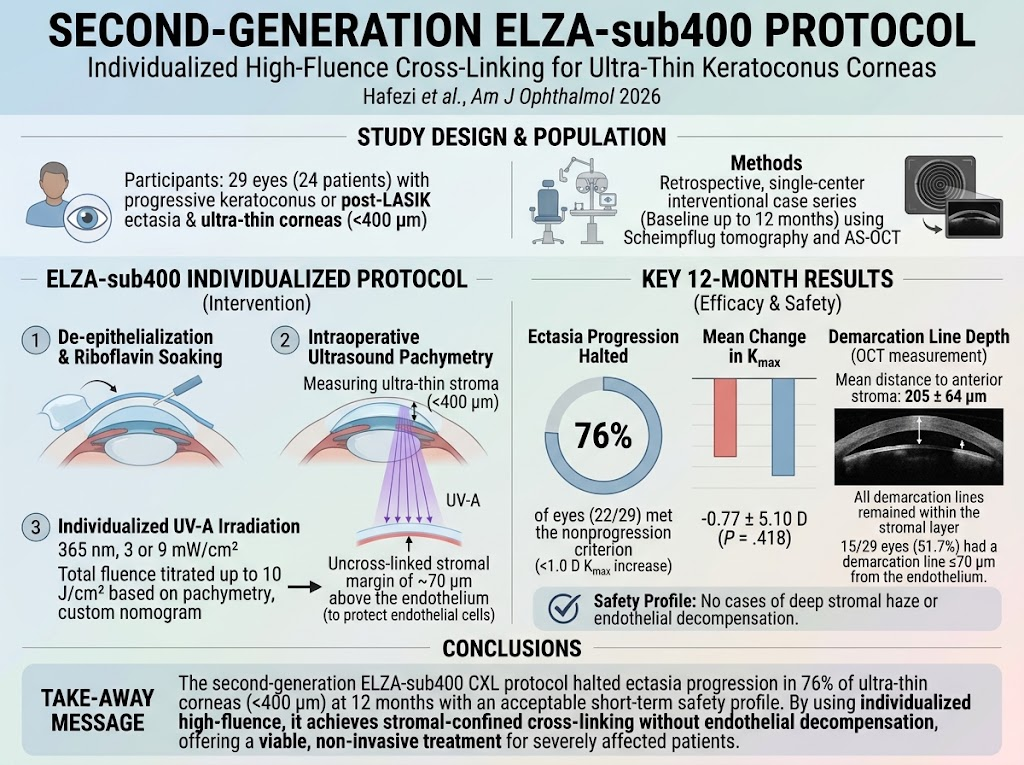
This retrospective, single-center, consecutive interventional case series included 29 eyes from 24 patients diagnosed with progressive keratoconus or post-LASIK ectasia, all featuring intraoperative stromal thickness under 400 µm measured by ultrasound pachymetry post riboflavin soaking and epithelium removal. The intervention consisted of an individualized, high-fluence continuous UV-A irradiation (365 nm) protocol using either 3 or 9 mW/cm². Total energy exposure was titrated up to 10 J/cm² following a previously published nomogram. This nomogram targeted maintaining an uncross-linked stromal margin of approximately 70 µm above the endothelium to safeguard endothelial integrity.
Main outcomes were assessed at baseline and through 12 months using corrected distance visual acuity (CDVA), corneal tomography from Scheimpflug imaging, anterior segment optical coherence tomography (AS-OCT), and Placido-based topography. The primary endpoint was defined as the proportion of eyes without keratoconus progression at 12 months, determined by less than 1.0 diopter increase in maximum keratometry (Kmax). Secondary measures included changes in visual acuity, refraction, corneal thickness, demarcation line depth, optical densitometry, and safety endpoints.
Key Findings
At 12 months, 76% (22/29) of eyes fulfilled the primary endpoint of disease nonprogression, with a mean change in Kmax of -0.77 ± 5.10 D—which was not statistically significant (P = .418). The demarcation line depth, a biomarker of cross-linking efficacy, averaged 205 ± 64 µm from the anterior stroma and 64 µm (IQR 49–152) from the endothelium, confirming targeted stromal penetration. Notably, all demarcation lines remained stromal, and 51.7% had a line within 70 µm of the endothelium, inline with the protocol’s margin goal.
Visual acuity outcomes showed a median CDVA change from 0.10 to 0.32 logMAR without statistical significance (P = .142). Minimal stromal thickness remained essentially stable with a median change of -4.0 µm (P = .309). Importantly, corneal densitometry measurements showed no significant increase, and no cases of deep stromal haze or endothelial decompensation were observed, supporting the short-term safety of the intervention.
These findings highlight the ELZA-sub400 protocol’s ability to deliver customized, high-fluence CXL safely to ultrathin corneas, achieving stromal strengthening without compromising the critical endothelial layer or inducing visually significant haze.
Expert Commentary
The ELZA-sub400 protocol exemplifies a precision medicine approach to CXL, leveraging intraoperative pachymetry and adjustable UV-A fluence to expand treatment eligibility to patients with advanced keratoconus who previously faced limited options. The ability to maintain an uncross-linked stromal safety margin is crucial in mitigating endothelial toxicity risks. The observed demarcation line characteristics indicate adequate stromal cross-linking depth, while the absence of endothelial complications confirms the protocol design’s protective efficacy.
Nonetheless, the cohort’s baseline severity and limited follow-up underline the need for larger, prospective studies to validate durability beyond one year and to clarify the impact on functional vision, given the numerically decreased CDVA observed. Furthermore, comparative studies against alternative ultrathin protocols or adjunct therapies could refine best practices.
Biological plausibility rests on UV-A energy dose-dependent collagen cross-link formation, which correlates with biomechanical reinforcement. Individualizing fluence based on real-time stromal thickness ensures therapeutic irradiation without exceeding cytotoxic thresholds for endothelial cells.
Conclusion
The second-generation ELZA-sub400 protocol represents a meaningful advancement in the management of ultrathin corneal ectasia, enabling individualized, high-fluence CXL that safely halts keratoconus progression in a substantial majority of patients at 12 months. While short-term safety and stromal handling are reassuring, the clinical significance of visual acuity changes and long-term outcomes require further elucidation in prospective, controlled investigations. This protocol has potential to reshape treatment paradigms for patients previously excluded from CXL due to tissue thinness, contributing to vision preservation in a vulnerable population.
Funding and Clinical Trials
The study does not specify funding sources or clinical trial registration information in the provided abstract. Future research should aim for transparency in trial registration and funding disclosures.
References
1. Hafezi F, Akcan RE, Kling S, et al. Second-Generation ELZA-sub400 Protocol: Individualized High-Fluence Cross-Linking for Ultra-Thin Keratoconus Corneas. Am J Ophthalmol. 2026 Jun 27;290:318-328. PMID: 42364705.
2. Wollensak G, Spoerl E, Seiler T. Riboflavin/ultraviolet-A-induced collagen crosslinking for the treatment of keratoconus. Am J Ophthalmol. 2003 May;135(5):620-7.
3. Kymionis GD, Diakonis VF, et al. Corneal cross-linking for ultrathin corneas. Surv Ophthalmol. 2014;59(4):420-8.
4. Raiskup-Wolf F, Hoyer A, et al. Corneal collagen crosslinking with riboflavin and ultraviolet-A light in keratoconus: long-term results. J Cataract Refract Surg. 2008 May;34(5):796-801.
5. Winn B, Hou Y, et al. Advances in corneal cross-linking techniques and technologies. Ophthalmol Clin North Am. 2013 Sep;26(3):481-94.