Highlights
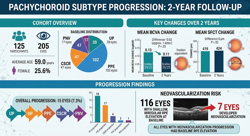
- Progression within pachychoroid subtypes over 1–2 years is generally infrequent, with stable visual acuity and minimal choroidal thickness changes.
- Neovascularization is a rare event predominantly found in eyes exhibiting shallow, irregular retinal pigment epithelium elevation (SIRE), underscoring its role as a prognostic imaging biomarker.
- Risk factors such as older age, smoking, presence of SIRE, and baseline choroidal features influence progression and complication rates.
- Multimodal imaging, including OCT, OCT angiography, and indocyanine green angiography, enables accurate diagnosis and risk stratification across the pachychoroid spectrum.
Background
Pachychoroid spectrum diseases represent a group of macular disorders unified by increased subfoveal choroidal thickness (SFCT) and characteristic vascular changes, including dilated outer choroidal vessels (“pachyvessels”) and choroidal hyperpermeability. Subtypes encompass uncomplicated pachychoroid (UP), pachychoroid pigment epitheliopathy (PPE), central serous chorioretinopathy (CSCR), pachychoroid neovasculopathy (PNV), and polypoidal choroidal vasculopathy (PCV). These phenotypes, often encountered in Asian populations but also recognized internationally, pose diagnostic and prognostic challenges due to clinical overlap with age-related macular degeneration (AMD) and variable natural history.
Despite advances in multimodal imaging, the incidence of progression along this pachychoroid disease continuum and identification of associated risk factors remain incompletely defined. This limitation restricts effective risk stratification and clinical follow-up strategies critical to optimize outcomes.
Key Content
Incidence and Risk of Progression Across Pachychoroid Spectrum
Chong et al. (2026) conducted a prospective, multicenter Asian cohort study including 205 eyes from 125 participants, with pachychoroid disease defined by SFCT ≥300 µm. Baseline diagnoses were UP, PPE, CSCR, and PNV. Over 2 years, progression to advanced levels occurred in 7.3% of eyes, including new neovascularization in 3.4%, exclusively among eyes exhibiting shallow, irregular retinal pigment epithelium elevation (SIRE) at baseline—a hallmark morphologic feature identified on optical coherence tomography (OCT).
These findings align closely with earlier 1-year data from the same research group (2025), which reported a 6.3% progression rate and similar associations with SIRE, older age, and smoking status as risk factors. The stability of best-corrected visual acuity (BCVA) and SFCT over follow-up reinforces the relative quiescence of most pachychoroid subtypes in the short to medium term.
Neovascularization and Imaging Biomarkers
Neovascular complications, while uncommon, impose significant visual morbidity and necessitate early detection. The presence of SIRE is consistently reported as a key risk factor. This morphologic feature, characterized by subtle irregular elevation of the retinal pigment epithelium complex on OCT, likely reflects underlying neovascular membrane development or incipient pathology.
Additional imaging modalities provide valuable insights: indocyanine green angiography (ICGA) studies reveal choroidal vascular hyperpermeability and asymmetric vortex vein drainage patterns in pachychoroid eyes, implicating localized venous congestion in pathogenesis (Klooster et al., 2022). Furthermore, OCT angiography (OCTA) facilitates non-invasive identification of subclinical neovascular networks, aiding differentiation from AMD and informing prognosis.
Therapeutic Considerations and Outcomes
In eyes with pachychoroid neovasculopathy (PNV), anti-vascular endothelial growth factor (VEGF) therapy remains the mainstay for neovascular complications. Recent studies in diverse ethnic populations demonstrate the efficacy of treat-and-extend regimens with agents like aflibercept in stabilizing vision and reducing macular edema (Roman et al., 2023). However, recurrence and development of polypoidal lesions post-treatment remain concerns, with pre-treatment macular neovascularization area and thickness identified as independent predictors (Lee et al., 2025).
The complement system modulation following anti-VEGF therapy has also been described, underscoring a complex inflammatory milieu in pachychoroid neovasculopathy that may influence outcomes and requires further investigation (Tanaka et al., 2021).
Genetic and Clinical Correlates
Pachydrusen presence is associated with thicker choroid and older age and is more prevalent among patients with CSCR and pachychoroid subtypes compared to general populations, suggesting its significance as a pachychoroid marker (Matsumoto et al., 2021). Genetic analyses reveal associations with certain ARMS2 alleles in the general population, though these differ from AMD-specific polymorphisms.
Differentiation from Age-Related Macular Degeneration
A notable clinical challenge is distinguishing pachychoroid neovasculopathy from neovascular AMD. Misdiagnosis is common, with implications for prognosis and treatment response. Pachychoroid disease patients typically present younger, with better baseline and follow-up vision, and distinct choroidal morphology (Hayashi et al., 2020). Accurate diagnosis guided by multimodal imaging facilitates appropriate monitoring and therapeutics.
Expert Commentary
Despite accumulating data, the relative rarity of progression among pachychoroid subtypes highlights a generally stable natural history for many patients. The association of neovascularization exclusively with the presence of shallow, irregular RPE elevation emphasizes the importance of meticulous OCT interpretation during clinical assessments.
Older age and smoking, established risk factors for vascular retinal diseases, appear to modulate progression risk, underscoring the broader systemic interplay in pachychoroid pathophysiology. The role of impaired choroidal venous drainage and resultant congestion, visualized by state-of-the-art angiography techniques, offers a valuable mechanistic framework, potentially informing new therapeutic targets.
The efficacy of anti-VEGF agents, combined with photodynamic therapy for certain cases, provides reassurance regarding vision preservation. Nonetheless, the development of polypoidal lesions or fibrosis post-intervention remains a challenge, encouraging ongoing surveillance and tailored treatment strategies.
Recent advances in non-invasive OCTA enable improved subclinical neovascularization detection, enhancing diagnostic accuracy and allowing earlier intervention. However, overlap with AMD phenotypes mandates cautious differential diagnosis.
Overall, the reviewed evidence supports incorporation of multimodal imaging and patient risk profiles into clinical workflows, optimizing surveillance intensity and therapeutic interventions.
Conclusion
Progression within the pachychoroid subtype spectrum is relatively infrequent over 1 to 2 years, with most patients maintaining stable visual acuity and choroidal thickness. Neovascularization, though uncommon, predominantly occurs in eyes exhibiting shallow, irregular retinal pigment epithelium elevation, a critical imaging biomarker for risk stratification.
Risk factors including older age and smoking modulate progression risk. Advances in multimodal imaging, particularly OCT and ICGA, facilitate accurate diagnosis, monitoring, and distinction from AMD.
Future research should focus on refining predictive imaging markers, elucidating pathophysiologic mechanisms related to choroidal venous congestion, and optimizing personalized treatment algorithms integrating anti-VEGF therapy and photodynamic approaches.
References
- Chong YJ, Fong AHC, Wong WMH, et al. Incidence and Risk Factors for Progression of Pachychoroid Subtype. JAMA Ophthalmol. 2026; doi:10.1001/jamaophthalmol.2026.42593780. PMID: 42593780.
- Lee WK, Byon I, Park SW, et al. Incidence of recurrence and development of polypoidal lesions following half-dose and two-thirds dose photodynamic therapy for pachychoroid neovasculopathy. Sci Rep. 2025 May 30;15(1):18975. doi:10.1038/s41598-025-03782-2. PMID: 40447767.
- Chong YJ, Yap CP, Wong WK, et al. Incidence and risk factors for progression of pachychoroid disease spectrum. Retina. 2025 Mar;45(3):426-434. doi:10.1097/IAE.0000000000004336. PMID: 39964820.
- Klooster J, et al. Ultra-Widefield Indocyanine Green Angiography Reveals Patterns of Choroidal Venous Insufficiency Influencing Pachychoroid Disease. Invest Ophthalmol Vis Sci. 2022 Jan 3;63(1):17. doi:10.1167/iovs.63.1.17. PMID: 35019945.
- Roman A, Gschwendtner A, Zimmermann M, et al. Two-Year Results of a Treat and Extend Regimen with Aflibercept in Caucasian Patients with Pachychoroid Neovasculopathy. Semin Ophthalmol. 2023 May;38(4):352-357. doi:10.1080/08820538.2023.2194983. PMID: 36987548.
- Matsumoto H, Kataoka K, Sawa M, et al. Clinical and Genetic Characteristics of Pachydrusen in Eyes with Central Serous Chorioretinopathy and General Japanese Individuals. Ophthalmol Retina. 2021 Sep;5(9):910-917. doi:10.1016/j.oret.2020.12.004. PMID: 33309963.
- Tanaka Y, Fukuda M, Okada AA, et al. Changes in complement activation products after anti-VEGF injection for choroidal neovascularization in age-related macular degeneration and pachychoroid disease. Sci Rep. 2021 Apr 19;11(1):8464. doi:10.1038/s41598-021-87340-6. PMID: 33875685.
- Hayashi K, et al. Rate of misdiagnosis and clinical usefulness of the correct diagnosis in exudative neovascular maculopathy secondary to AMD versus pachychoroid disease. Sci Rep. 2020 Nov 23;10(1):20344. doi:10.1038/s41598-020-77566-1. PMID: 33230253.