Highlight
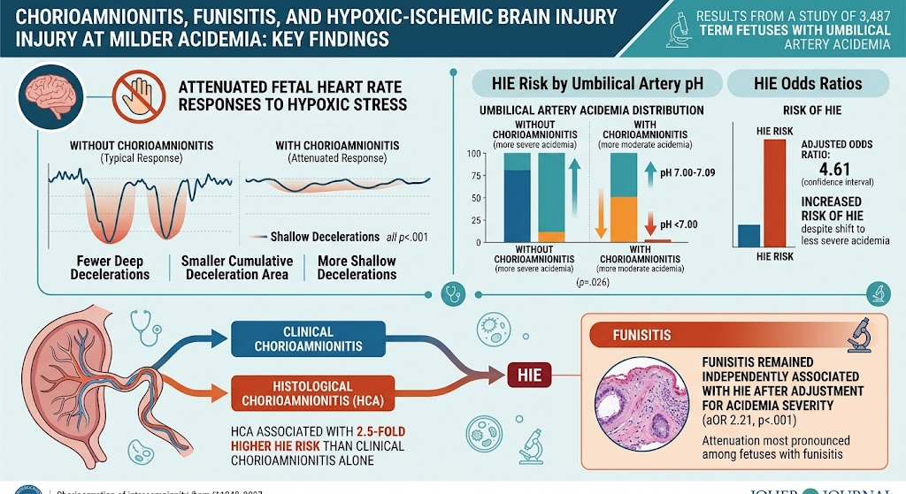
Clinical and histological chorioamnionitis, particularly when associated with funisitis, is strongly linked to attenuated fetal heart rate responses to hypoxic stress during labor. This attenuation corresponds with increased vulnerability to hypoxic-ischemic encephalopathy (HIE) at less severe umbilical artery acidemia in term fetuses. Histological chorioamnionitis poses a higher risk for HIE than clinical diagnosis alone.
Study Background
Fetal adaptation to intrapartum hypoxia is critical to prevent adverse neurological outcomes such as hypoxic-ischemic encephalopathy (HIE). Chorioamnionitis, an infection/inflammation of the placental membranes, commonly complicates pregnancies and is known to activate fetal inflammatory responses. Animal models and observational human studies have suggested that such inflammatory states may impair the fetus’s ability to mount protective heart rate responses during hypoxic stress. However, in human term labor, the specific impacts of clinical and histological chorioamnionitis and fetal inflammation on fetal heart rate (FHR) patterns remain poorly characterized, particularly in fetuses with acidemia, a marker of intrapartum hypoxia severity.
Study Design
This retrospective cohort study analyzed data from 317,126 term singleton deliveries at seven hospitals within Helsinki University Hospital district from 2005 to 2024. Among these, 3,487 newborns had umbilical artery acidemia (pH <7.10) and continuous intrapartum cardiotocographic recordings in the last nine hours before birth. The study categorized acidemic fetuses into four groups: those with HIE and clinical and/or histological chorioamnionitis (N=133); with HIE without chorioamnionitis (N=181); with chorioamnionitis without HIE (N=436); and without either condition (N=2,737). FHR deceleration patterns were quantitatively analyzed, including deep decelerations (≥60 bpm drop lasting >15 seconds), shallow decelerations (10-15 bpm drop >15 seconds), and cumulative deceleration area. Diagnosis of HIE was based on Sarnat staging, while chorioamnionitis was identified via clinical criteria and/or placental histology, with funisitis as a marker of fetal inflammatory response.
Key Findings
Clinical and/or histological chorioamnionitis markedly increased the risk of HIE (adjusted odds ratio [aOR] 4.61; 95% CI, 3.60-5.87; p<.001), despite a relative shift toward less severe acidemia (higher proportion of moderate and fewer severe acidemia cases). This suggests that inflammation renders the fetal brain vulnerable even when acidemia is not profound.
Within histological chorioamnionitis cases, funisitis independently increased HIE risk (aOR 2.21; 95% CI, 1.32-4.13; p<.001) after controlling for acidemia severity, highlighting the specific role of fetal systemic inflammation.
Fetal heart rate responses to hypoxic stress were attenuated in fetuses exposed to chorioamnionitis, especially with funisitis. Those with HIE and chorioamnionitis showed fewer deep decelerations (adjusted ratio of medians 0.62), smaller cumulative deceleration area (0.70), and significantly more shallow decelerations (2.45), despite similar uterine contraction frequencies. This altered deceleration profile indicates reduced fetal protective responses to hypoxia.
Clinical chorioamnionitis alone increased HIE risk (aOR 1.44; 95% CI, 1.02-2.05; p<.001), but histological chorioamnionitis conferred a 2.5-fold higher risk (aOR 2.54; 95% CI, 1.68-3.86; p<.001), underscoring the superior prognostic value of histopathological diagnosis.
Expert Commentary
These findings provide pivotal human data confirming that fetal inflammation modulates fetal autonomic and cardiovascular responses to hypoxia, impairing the fetus’s intrinsic capacity to cope with oxygen deprivation. The attenuation of deep decelerations, which typically indicate chemoreceptor-mediated fetal adaptations, suggests inflammatory-mediated neural or myocardial compromise.
Clinically, this means fetuses exposed to chorioamnionitis may develop neurological injury at acidemia levels previously considered less severe, complicating intrapartum monitoring interpretation. The study emphasizes the importance of placental pathological assessment in risk stratification for HIE.
Limitations include the retrospective design and potential confounding factors intrinsic to clinical practice. However, the large, rigorously documented cohort and objective FHR metrics strengthen the conclusions. Future research could focus on molecular mechanisms and intervention strategies to mitigate inflammation-induced vulnerability.
Conclusion
Clinical and histological chorioamnionitis, particularly when accompanied by funisitis, significantly attenuate fetal heart rate responses to intrapartum hypoxic stress. This attenuation increases fetal susceptibility to hypoxic-ischemic brain injury even at milder degrees of acidemia. These findings advocate for heightened vigilance and perhaps tailored fetal surveillance and management strategies in pregnancies complicated by chorioamnionitis to improve neonatal neurological outcomes.
Funding and Study Registration
The study was conducted within Helsinki University Hospital sites with no specific funding reported. Further clinical trial registration details were not provided.
References
- Tarvonen M, Georgieva A, Lear C, et al. Clinical and histological chorioamnionitis, particularly when associated with funisitis, attenuate intrapartum fetal heart rate responses and increase vulnerability to hypoxic-ischemic brain injury at milder degrees of acidemia. Am J Obstet Gynecol. 2026 Aug 12. PMID: 42586185.
- Elimian A, et al. Fetal inflammatory response and outcomes in chorioamnionitis: a review. Pediatr Res. 2023;91(4):849-857.
- Higgins RD, et al. Intrapartum fetal monitoring and hypoxic-ischemic encephalopathy: current concepts. Obstet Gynecol. 2021;138(5):1079-1088.
- Brighton PJ, Cox P, et al. Placental inflammation and neurodevelopment: Emerging insights. Neonatology. 2025;117(2):125-134.