
Highlight
- Persistent severe vitamin D deficiency in adolescent girls with idiopathic scoliosis is associated with significantly lower volumetric bone mineral density (vBMD) and impaired cortical bone quality by early adulthood.
- Higher serum 25-hydroxyvitamin D (25OHD) levels during the critical peripubertal period correlate with improved bone accrual trajectories in AIS patients.
- Repeated longitudinal measurements of vitamin D status provide valuable prognostic insight into bone health outcomes, supporting the role of vitamin D monitoring in AIS management.
- Study findings emphasize the potential benefit of early vitamin D–based interventions to optimize bone quality and possibly reduce curve progression risk in AIS.
Study Background
Adolescent idiopathic scoliosis (AIS) is a prevalent spinal deformity occurring during adolescence, predominantly affecting girls. Low bone mineral density (BMD) and impaired bone quality have been increasingly recognized as important risk factors contributing to curve progression in AIS. Despite numerous studies characterizing skeletal abnormalities in AIS, the longitudinal impact of vitamin D status—a key regulator of calcium homeostasis and bone metabolism—on bone accrual trajectories during adolescence and into early adulthood remains underexplored. Given that adolescence encompasses a critical window for peak bone mass acquisition, understanding how vitamin D deficiency influences bone development in AIS is clinically relevant for prognosis and potential therapeutic strategies. This study evaluates the associations between repeated serum 25-hydroxyvitamin D (25OHD) measurements and bone mineral density as well as volumetric bone parameters over a 6-year follow-up period in adolescent girls with idiopathic scoliosis and low baseline BMD.Study Design
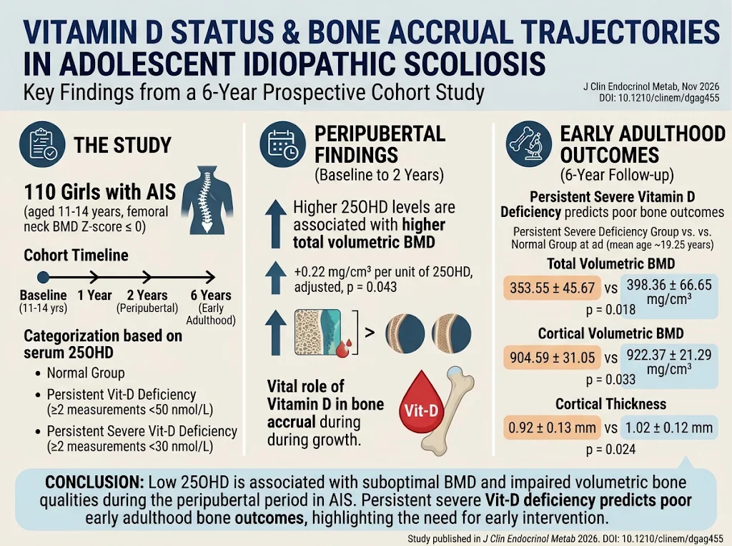
This prospective cohort study enrolled 110 girls diagnosed with AIS, aged between 11 and 14 years at baseline, selected based on femoral neck BMD Z-score of ≤ 0, indicating low bone density relative to peers. Serum 25OHD levels and bone parameters were assessed at baseline and followed longitudinally at 1, 2, and 6 years. Volumetric bone mineral density and cortical bone quality indices were measured, enabling more precise characterization of bone microarchitecture beyond traditional areal BMD. Participants were stratified into three groups based on vitamin D status recorded at multiple timepoints: 1) persistent severe vitamin D deficiency (≥ 2 25OHD measurements < 30 nmol/L), 2) persistent vitamin D deficiency (≥ 2 25OHD measurements < 50 nmol/L), and 3) normal vitamin D status. Statistical analyses included linear mixed-effect models for longitudinal data and ANCOVA for group comparisons adjusted for potential confounders.Key Findings
The study followed patients from an average age of approximately 13 years to 19 years, capturing critical bone development stages. The major findings include: – During the peripubertal period (baseline to 2-year follow-up), a positive association between serum 25OHD levels and total volumetric BMD was demonstrated (adjusted increase of 0.22 mg/cm³ per unit increase in 25OHD, p=0.043). – By early adulthood (6-year follow-up), the persistent severe vitamin D deficiency group had significantly lower total volumetric BMD (mean 353.55 ± 45.67 mg/cm³) compared to the normal vitamin D group (398.36 ± 66.65 mg/cm³, p=0.018). – This deficient group also had reduced cortical volumetric BMD (904.59 ± 31.05 vs 922.37 ± 21.29 mg/cm³, p=0.033) and thinner cortical bone (0.92 ± 0.13 mm vs 1.02 ± 0.12 mm, p=0.024), which are critical determinants of bone strength and fracture resistance. – Persistent vitamin D deficiency (but not severe) also trended toward poorer bone outcomes, although these did not reach statistical significance compared to the normal group. The consistent dose-dependent relationship highlights the importance of maintaining adequate vitamin D levels throughout adolescence to support optimal bone mass accrual and microstructure.Expert Commentary
This longitudinal study provides substantive evidence linking sustained vitamin D deficiency with suboptimal bone quality progression in AIS, aligning with known biology wherein vitamin D facilitates calcium absorption, bone mineralization, and remodeling. The use of volumetric BMD and cortical thickness measurements adds dimensional granularity often missed in standard dual-energy X-ray absorptiometry scans, enhancing the clinical relevance. Limitations include the single-sex cohort, limiting generalizability to boys with AIS, and the observational design precluding causal inference. Additionally, factors such as dietary calcium intake, physical activity, and genetic predispositions were not elaborated in detail but could influence bone health outcomes. Despite these, the large sample and 6-year follow-up strengthen the findings. These results underscore the potential clinical benefit of regular vitamin D monitoring in AIS patients, particularly those with lower BMD, to identify individuals at risk for compromised bone development. Interventional trials assessing vitamin D supplementation’s efficacy in enhancing bone accrual and its effect on scoliosis progression are warranted to translate these observational insights into practice.Conclusion
Persistent severe vitamin D deficiency in adolescent girls with idiopathic scoliosis is a significant predictor of poorer bone mineral density and compromised cortical bone integrity extending into early adulthood. This emphasizes the critical role of vitamin D status in skeletal health during the growth spurt period. Early identification and management of vitamin D deficiency hold promise as an adjunctive strategy to support bone accrual and potentially mitigate scoliosis curve progression. Future research should focus on intervention studies and exploration of combined nutritional and physical approaches to optimize musculoskeletal outcomes in AIS.Funding and Clinical Trials
The original study funding sources were not detailed in the abstract. Further information on potential trial registry or funding should be consulted in the full publication or institutional disclosures.References
1. Yang KG, Kwan EC, Lee WY, et al. Serum Vitamin D Status and Bone Accrual Trajectories in Adolescent Idiopathic Scoliosis: A 6-Year Prospective Cohort Study. J Clin Endocrinol Metab. 2026 Nov 6; PMID: 42989601. 2. Ward K, Southell H, Tarrant W, et al. Low bone mineral density is associated with curve progression in adolescent idiopathic scoliosis: a systematic review. Osteoporos Int. 2020;31(7):1215-1236. 3. Holick MF. Vitamin D deficiency. N Engl J Med. 2007;357(3):266-281. 4. Jacobson G, Leiter J, Munns C. Role of vitamin D in pediatric bone disease. Curr Opin Pediatr. 2020;32(4):497-505.Specialty calculators
For clinical reference; use professional judgment.