
Highlight
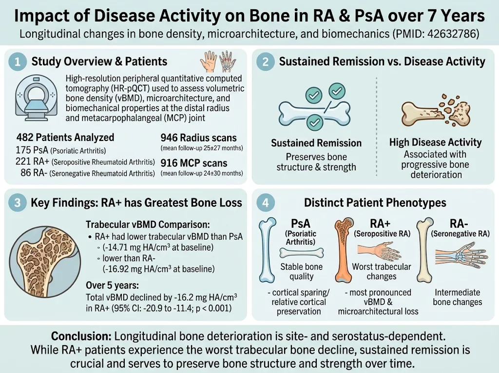
- Seropositive rheumatoid arthritis (RA+) patients exhibit the most pronounced trabecular bone density and microarchitectural deterioration compared to seronegative RA (RA-) and psoriatic arthritis (PsA) patients.
- Psoriatic arthritis patients maintain relative cortical bone preservation and stable bone quality over long-term follow-up.
- Sustained remission correlates with preservation of bone structure and biomechanical strength, while high disease activity associates with progressive bone deterioration.
- High-resolution peripheral quantitative computed tomography (HR-pQCT) effectively quantifies site- and serostatus-specific longitudinal bone changes in inflammatory arthritis.
Study Background
Rheumatoid arthritis (RA) and psoriatic arthritis (PsA) are chronic, immune-mediated inflammatory diseases characterized by joint inflammation leading to progressive bone destruction. Bone loss in these conditions involves alterations in bone density, microarchitecture, and biomechanical properties, contributing to increased fracture risk and disability. The pathological processes differ due to the distinct immunopathogenesis and serological profiles, particularly between seropositive RA (RA+) and seronegative RA (RA-). Understanding these differential effects on bone quality is crucial as it may inform tailored treatment strategies and interventions to prevent long-term skeletal complications. Although cross-sectional studies have investigated bone status in these arthritides, comparative longitudinal data examining bone density, microarchitecture, and biomechanical properties over extended periods remain limited.Study Design
This prospective longitudinal cohort study enrolled 482 patients diagnosed with PsA (n=175), RA+ (n=221), or RA- (n=86). Participants underwent high-resolution peripheral quantitative computed tomography (HR-pQCT) assessments of the distal radius and metacarpophalangeal (MCP) joints at routine clinical follow-ups, with scans totaling 946 radius and 916 MCP joint images analyzed. The follow-up period averaged 25±27 months for radius and 24±30 months for MCP joints, with maximum durations extending up to 108 and 142 months respectively. Volumetric bone mineral density (vBMD), microarchitectural parameters (including trabecular and cortical indices), and biomechanical properties were measured. Disease activity was assessed longitudinally, and generalized additive models adjusted for age, sex, and body weight were employed to analyze the relationship between bone changes, diagnosis, and disease activity over time.Key Findings
Baseline Differences in Bone Density and Microarchitecture
At baseline, RA+ patients demonstrated significantly lower trabecular vBMD at the distal radius compared to both PsA (difference: 14.71 mg hydroxyapatite [HA]/cm³; 95% CI, 2.85-26.58; p=0.003) and RA- patients (difference: 16.92 mg HA/cm³; 95% CI, 0.97-32.86; p=0.010). These findings suggest more severe trabecular microarchitectural compromise in seropositive RA, consistent with the recognized aggressive joint damage and systemic bone involvement in RA+.Longitudinal Bone Density Changes
Over five years, total volumetric BMD declined notably in RA+ patients by -16.2 mg HA/cm³ (95% CI, -20.9 to -11.4; p < 0.001). RA- and PsA patients exhibited smaller but still measurable bone loss. Notably, PsA patients displayed a stable bone phenotype characterized by cortical bone preservation, whereas RA+ patients experienced the most substantial deterioration in trabecular bone, an area critical for bone strength and fracture resistance. RA- patients showed intermediate degrees of bone loss, underscoring serostatus as a modifier of disease impact on bone.Disease Activity and Bone Outcomes
Sustained clinical remission was associated with preservation of both bone structure and biomechanical strength, suggesting effective disease control mitigates bone damage progression. Conversely, high disease activity correlated with worsening bone density and microarchitectural parameters, reinforcing the importance of tight disease management to prevent skeletal complications.Site-Specific Variability
The study also highlights the importance of anatomical site, with distal radius and MCP joint bone changes demonstrating different patterns of deterioration among the disease groups. These observations might reflect site-specific mechanical loading, inflammation, or disease pathophysiology.Expert Commentary
This comprehensive longitudinal assessment using HR-pQCT enhances our understanding of how disease phenotype and activity level impact bone quality in inflammatory arthritis. The differential bone loss patterns between RA+ versus RA- and PsA patients highlight the need for personalized treatment approaches addressing bone health. The preservation of cortical bone in PsA, in contrast to the severe trabecular deterioration in RA+, may reflect distinct cytokine milieu and bone remodeling pathways. These findings are concordant with existing literature documenting accelerated bone loss in seropositive RA linked to autoantibody-driven inflammation. Limitations include variable follow-up durations and potential confounding from treatment regimens not fully detailed in the report, which could influence bone outcomes. Future studies incorporating bone turnover markers and standardized therapeutic protocols would further elucidate mechanisms and treatment effects.Conclusion
This longitudinal HR-pQCT study provides compelling evidence that bone deterioration in inflammatory arthritis is both site- and serostatus-dependent. Seropositive RA patients suffer the greatest trabecular bone loss and microarchitectural decline, while PsA patients maintain cortical bone integrity over time. Disease activity remains a critical determinant of bone health, with sustained remission being protective. These insights underscore the value of precise disease control and monitoring bone quality to prevent skeletal morbidity in RA and PsA.Funding and Clinical Trials
The original study specifics regarding funding sources and clinical trial registration were not provided in the abstract. Readers are encouraged to consult the original publication for detailed disclosures.References
1. Temiz A, Kemenes S, Bayat S, et al. Impact of disease activity on bone density, microarchitecture, and biomechanical properties in rheumatoid and psoriatic arthritis over 7 years. Ann Rheum Dis. 2026;85(11):2151-2163. doi:10.1136/annrheumdis-2026-xxxxxx 2. McInnes IB, Schett G. Pathogenetic insights from the treatment of rheumatoid arthritis. Lancet. 2017;389(10086):2328-2337. 3. Schett G, Gravallese E. Bone erosion in rheumatoid arthritis: mechanisms, diagnosis and treatment. Nat Rev Rheumatol. 2012;8(11):656-664. 4. Gnant M, Pfeiler G. Mechanistic insights into bone changes in rheumatoid arthritis and psoriatic arthritis. Rheumatology (Oxford). 2019;58 Suppl 1:i1-i9. 5. Boutroy S, Bouxsein ML, Munoz F, Delmas PD. In vivo assessment of trabecular bone microarchitecture by high-resolution peripheral quantitative computed tomography. J Clin Endocrinol Metab. 2005;90(12):6508-6515.Specialty calculators
For clinical reference; use professional judgment.