Highlight
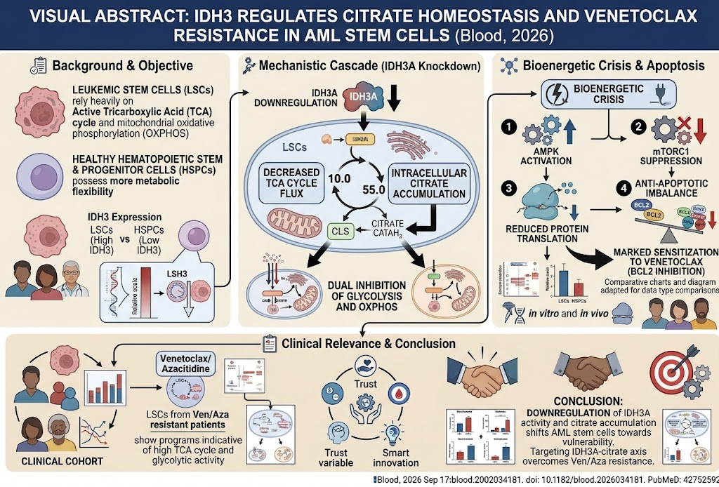
– IDH3A is a selective regulator of leukemic stem cell (LSC) maintenance in acute myeloid leukemia (AML).
– IDH3A knockdown disrupts TCA cycle flux, causing citrate accumulation and bioenergetic crisis in LSCs but spares healthy hematopoietic stem cells.
– Metabolic perturbation through IDH3A downregulation sensitizes AML LSCs to the BCL2 inhibitor venetoclax.
– Venetoclax-resistant patient-derived AML LSCs exhibit elevated TCA cycle and glycolytic activity, linked to IDH3-mediated metabolic adaptation.
Study Background
Acute myeloid leukemia is a heterogeneous hematologic malignancy characterized by clonal expansion of undifferentiated myeloid blasts. Despite advances in therapy, disease relapse remains frequent, largely due to persistence of leukemic stem cells (LSCs) which exhibit distinct metabolic dependencies, including reliance on mitochondrial oxidative phosphorylation and the tricarboxylic acid (TCA) cycle. Identification of metabolic vulnerabilities specific to LSCs could facilitate development of targeted therapies that overcome treatment resistance, particularly resistance to BCL2 inhibitor venetoclax combined with hypomethylating agents such as azacitidine (Ven/Aza). The metabolic enzyme isocitrate dehydrogenase 3 (IDH3) emerged as a potential key regulator in this context.
Study Design
The study conducted by Demir et al. employed a comprehensive multi-model approach, integrating transcriptomic profiling, in vitro assays, and in vivo transplantation models to dissect the role of IDH3, specifically the catalytic subunit IDH3A, in AML LSCs versus healthy hematopoietic stem and progenitor cells (HSPCs). Patient-derived AML samples including those from individuals exhibiting Ven/Aza resistance were analyzed to correlate metabolic gene signatures with clinical outcomes. Functional validation involved shRNA-mediated knockdown of IDH3A in AML cells to assess changes in colony formation, bone marrow organoid growth, and engraftment potential in xenograft models, alongside metabolic flux analysis and biochemical assays to measure intracellular citrate, glycolytic, and oxidative phosphorylation activity. Venetoclax sensitivity assays were conducted both in vitro and in vivo.
Key Findings
IDH3A Expression and Clinical Correlation: IDH3A protein levels were significantly higher in LSCs compared to healthy HSPCs. Transcriptomic data from AML patient cohorts revealed a positive correlation between heightened TCA cycle activity, mediated via IDH3, and poorer clinical outcomes, including resistance to venetoclax-based therapies.
Functional Impact of IDH3A Knockdown: Targeted suppression of IDH3A impaired AML LSC maintenance by reducing colony-forming capacity and compromising growth within a bone marrow organoid model. Critically, healthy hematopoiesis was minimally affected, underscoring a therapeutic window. In xenotransplantation models, IDH3A knockdown reduced in vivo LSC engraftment and leukemia propagation.
Metabolic Consequences of IDH3A Deficiency: Depletion of IDH3A resulted in the accumulation of intracellular citrate due to attenuation of TCA cycle flux. This metabolic bottleneck concomitantly suppressed both glycolysis and mitochondrial oxidative phosphorylation, triggering a bioenergetic crisis in AML LSCs. Energy deprivation activated the AMP-activated protein kinase (AMPK) pathway and inhibited mammalian target of rapamycin complex 1 (mTORC1), leading to reduced protein synthesis and altered expression of anti-apoptotic proteins.
Modulation of Venetoclax Sensitivity: The metabolic reprogramming induced by IDH3A knockdown sensitized AML LSCs to BCL2 inhibition with venetoclax, demonstrated by enhanced cell death both in vitro and in vivo. Notably, AML LSCs isolated from patients with clinical resistance to Ven/Aza displayed transcriptomic signatures indicative of elevated TCA cycle and glycolytic activity, consistent with a metabolically active state that IDH3 targeting could disrupt.
Expert Commentary
This study highlights IDH3A as a pivotal metabolic rheostat in AML LSCs, sustaining their bioenergetic fitness and contributing to therapy resistance. The distinction between metabolically inflexible LSCs and more adaptable healthy HSPCs reinforces the therapeutic selectivity potential. Mechanistically, the interplay between citrate homeostasis, metabolic flux, and apoptotic regulation underscores the intricate cross-talk linking metabolism to cell survival pathways. Targeting IDH3A or its metabolic axis may offer a novel approach to overcome the limitations of current venetoclax-based regimens, especially in refractory AML.
Limitations of the study include the need for elaboration on potential off-target effects of IDH3A knockdown and evaluation of combinatory strategies in clinical contexts. Moreover, IDH3 mutations and variants in AML—distinct from IDH1/2 mutations—need further characterization to determine their broader relevance.
Conclusion
IDH3A functions as a critical metabolic regulator that maintains TCA cycle activity and energy homeostasis in AML LSCs. Its inhibition induces citrate accumulation and a metabolic crisis that impairs leukemic stem cell function and heightens vulnerability to venetoclax. These findings support the IDH3A-citrate axis as a promising target to overcome venetoclax resistance and improve treatment outcomes in AML patients, warranting further preclinical validation and clinical exploration.
Funding and Clinical Trials
Information on funding sources and clinical trial registrations was not detailed in the primary publication. Future clinical studies are necessary to evaluate the translational potential of IDH3 targeting strategies in AML.
References
Demir A, Mönnig M, Aroua N, et al. IDH3 regulates citrate homeostasis to control metabolic fitness and venetoclax resistance of AML stem cells. Blood. 2026 Sep 17. PMID: 42752592. https://pubmed.ncbi.nlm.nih.gov/42752592/