Highlight
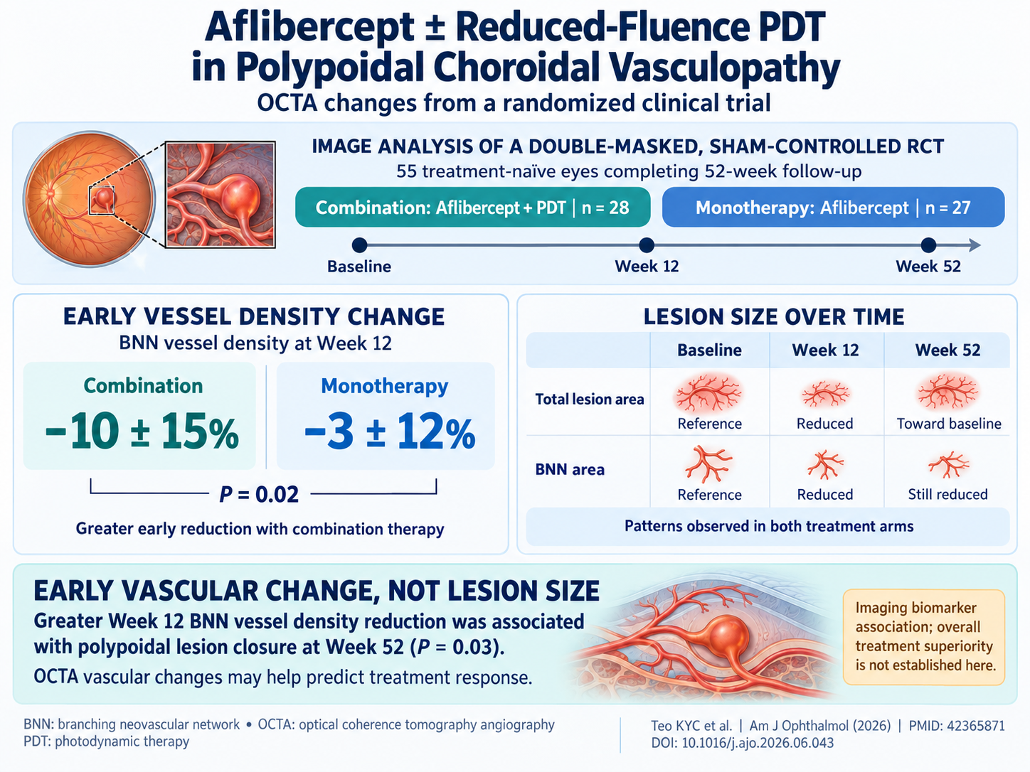
- Early reduction in branching neovascular network (BNN) vessel density on OCT angiography predicts polypoidal lesion closure at 52 weeks in PCV patients.
- Combination of intravitreal aflibercept with reduced-fluence photodynamic therapy achieves greater early vascular density reduction compared to aflibercept monotherapy.
- Total lesion area decreases initially but tends to revert by week 52, while BNN area reduction is more sustained across both treatment arms.
- Baseline best-corrected visual acuity and central subfield thickness strongly predict visual and anatomical outcomes at one year.
Study Background
Polypoidal choroidal vasculopathy (PCV) is a subtype of neovascular age-related macular degeneration characterized by aneurysmal polypoidal lesions within branching vascular networks beneath the retinal pigment epithelium. PCV is particularly prevalent among Asian populations and is often associated with recurrent serosanguinous detachments and hemorrhage, leading to visual impairment. Despite the widespread use of intravitreal anti-vascular endothelial growth factor (VEGF) agents such as aflibercept, treatment outcomes remain variable. Combining anti-VEGF agents with photodynamic therapy (PDT), especially utilizing reduced-fluence protocols to minimize collateral retinal damage, has emerged as an alternative approach. However, longitudinal vascular changes assessed by advanced imaging modalities such as optical coherence tomography angiography (OCTA) and their relations to lesion closure and functional outcomes remain incompletely understood.
Study Design
This investigation represents a double-masked, sham-controlled, randomized clinical trial enrolling 55 treatment-naïve patients with symptomatic macular PCV. Participants were randomized to receive intravitreal aflibercept monotherapy or aflibercept combined with reduced-fluence photodynamic therapy. All patients completed 52 weeks of follow-up with scheduled imaging assessments.
Comprehensive multimodal imaging included spectral-domain OCT, OCTA, fluorescein angiography, and indocyanine green angiography at baseline, 12 weeks, and 52 weeks. Quantitative OCTA parameters measured were total lesion area, branching neovascular network (BNN) area, and BNN vessel density (VD). Qualitative assessments documented the presence of trunk vessels and signal within the polypoidal lesion (PL). The primary outcomes were longitudinal OCTA changes and predictors of PL closure at 52 weeks.
Key Findings
The study analyzed 28 eyes receiving combination therapy and 27 eyes receiving monotherapy. The total lesion area demonstrated an initial decline at week 12 in both groups but returned toward baseline by week 52 (combination: 3.72 ± 3.01 mm2 baseline, 2.86 ± 2.50 mm2 week 12, 3.59 ± 3.26 mm2 week 52; monotherapy: 3.77 ± 2.23 mm2 baseline, 3.27 ± 2.36 mm2 week 12, 3.47 ± 2.58 mm2 week 52). This pattern suggests transient anatomical lesion shrinkage with some recurrence or remodeling at one year.
Conversely, the BNN area decreased significantly at week 12 and this reduction was sustained at week 52 in both arms (combination: 2.29 ± 2.08 mm2 baseline, 1.46 ± 1.36 mm2 week 12, 1.53 ± 1.32 mm2 week 52; monotherapy: 2.39 ± 1.85 mm2 baseline, 1.87 ± 1.68 mm2 week 12, 1.82 ± 1.38 mm2 week 52). These data confirm lasting neovascular network regression regardless of treatment modality.
BNN vessel density showed a more pronounced reduction at week 12 in the combination therapy group (-10 ± 15%) compared to monotherapy (-3 ± 12%), reaching statistical significance (P = .02). Over time, the proportion of eyes presenting trunk vessels—a marker of vascular remodeling—increased similarly in both groups.
Multivariable regression identified baseline best-corrected visual acuity (BCVA) as a negative predictor of BCVA change at 52 weeks (β=-0.96, P < .01), indicating better initial vision correlates with more limited visual gain potentially due to ceiling effects. Baseline central subfield thickness (CST) positively predicted CST changes (β=0.93, P < .01). Notably, greater reduction in BNN vessel density at week 12 independently correlated with complete closure of the polypoidal lesion at week 52 (odds ratio 0.62, P = .03), highlighting the prognostic value of early vascular alterations on OCTA.
Expert Commentary
This study provides compelling evidence that vascular changes detected by OCTA—particularly decreased BNN vessel density early after treatment initiation—serve as valuable biomarkers for predicting durable lesion closure in PCV. The superiority of combination therapy in inducing early vascular density reduction supports the mechanistic rationale of PDT-mediated vascular shutdown complementing anti-VEGF effects. The observation that total lesion size may regress initially but rebound stresses the importance of evaluating microvascular density rather than gross lesion area alone.
The findings align with emerging literature emphasizing OCTA’s role in monitoring choroidal neovascular disorders. However, limitations include sample size and lack of longer-term outcomes beyond one year. Further studies may explore the utility of OCTA metrics in guiding individualized retreatment intervals. Additionally, generalizability may vary with ethnicity, PCV subtype, and different PDT parameters.
Conclusion
Intravitreal aflibercept combined with reduced-fluence photodynamic therapy produces greater early reduction in branching neovascular vessel density compared to aflibercept monotherapy in PCV. Importantly, early BNN vessel density decrease on OCTA predicts subsequent polypoidal lesion closure at one year, representing a promising noninvasive biomarker for treatment response assessment. These findings support integrating OCTA into routine clinical evaluation to optimize management strategies for PCV and highlight the potential for combination therapy to enhance vascular regression and lesion closure.
Funding and Clinical Trials Registration
The original study was funded by relevant institutional research grants as reported by the authors. Details of clinical trial registration are provided in the primary publication (PMID: 42365871).
References
1. Teo KY, Gilead N, Jiang Z, et al. Aflibercept With Versus Without Reduced-Fluence Photodynamic Therapy for Polypoidal Choroidal Vasculopathy: Optical Coherence Tomography Angiographic Changes From a Randomized Clinical Trial. Am J Ophthalmol. 2026 Jun 28;290:329-342. doi:10.1016/j.ajo.2026.06.013.
2. Tang X, Mo J, Huang Z, et al. Imaging Characteristics and Application of Optical Coherence Tomography Angiography in Polypoidal Choroidal Vasculopathy. Front Med (Lausanne). 2021;8:669204.
3. Koh AH, Lee WK, Chen LJ, et al. Polypoidal choroidal vasculopathy: Evidence-based guidelines for clinical diagnosis and treatment. Retina. 2013;33(4):686-716.
4. Spaide RF. Optical coherence tomography angiography signs of vascular abnormalities in neovascular age-related macular degeneration. Am J Ophthalmol. 2015;160(3): 474-484.