Highlight
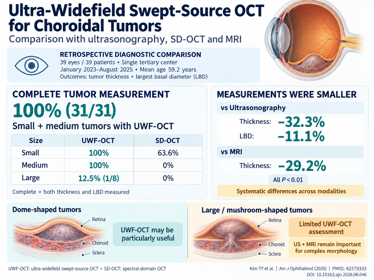
- Ultra-widefield swept-source OCT (UWF-OCT) achieves 100% successful measurement of thickness and basal diameter in small-to-medium choroidal tumors, outperforming spectral-domain OCT.
- UWF-OCT measurements tend to underestimate tumor size compared to ultrasonography (US) and magnetic resonance imaging (MRI), reflecting its detail-oriented but limited penetration depth.
- Mushroom-shaped tumor morphology significantly reduces UWF-OCT image quality, limiting applicability in large or complex tumors.
- Multimodal imaging remains necessary for comprehensive evaluation, yet UWF-OCT offers precise, noninvasive assessment valuable for diagnosis and longitudinal monitoring.
Study Background and Disease Burden
Choroidal tumors, including melanomas, metastatic lesions, hemangiomas, and osteomas, represent a significant clinical challenge due to their potential for vision loss and metastasis, alongside diagnostic difficulty. Accurate measurement of tumor dimensions, especially thickness and largest basal diameter (LBD), is crucial for diagnosis, treatment planning, and monitoring. Conventional imaging modalities such as ultrasonography and magnetic resonance imaging (MRI) provide critical information but have limitations in resolution, invasiveness, or accessibility. Spectral-domain optical coherence tomography (SD-OCT) offers fine structural imaging but is often restricted by a limited field of view and penetration depth, especially for tumors beyond small size. The advent of ultra-widefield swept-source OCT (UWF-OCT), a technology capable of deeper and broader ocular imaging, promises enhanced clinical utility but requires rigorous evaluation against established modalities. This study addresses this gap, aiming to define UWF-OCT’s role in the clinical assessment of intraocular tumors.
Study Design
This retrospective diagnostic comparison included 39 eyes from 39 patients diagnosed with choroidal tumors at a single tertiary referral center between January 2023 and August 2025. The tumor types were predominantly choroidal melanomas (69.2%), with metastatic tumors (12.8%), hemangiomas (10.3%), osteomas (5.1%), and indeterminate melanocytic lesions (2.6%). All patients underwent imaging by ultra-widefield swept-source OCT, ultrasonography (US), spectral-domain OCT (SD-OCT), and magnetic resonance imaging (MRI) at diagnosis. The primary endpoints were tumor thickness and largest basal diameter (LBD) measurements across modalities, as well as the measurability rate in different tumor size categories (small, medium, large). Data analysis included comparative statistics and logistic regression to identify predictive factors influencing UWF-OCT applicability.
Key Findings and Results
UWF-OCT demonstrated unparalleled capability in measuring tumor dimensions in small and medium choroidal tumors. It successfully obtained complete thickness and LBD measurements in 100% (31/31) of these tumors, substantially outperforming SD-OCT, which achieved complete measurements in only 63.6% of small tumors and failed to measure medium or large tumors completely.
When comparing dimensional outputs, UWF-OCT consistently reported smaller tumor thickness (-32.3%) and LBD (-11.1%) than ultrasonography (P < 0.01 for both) and similarly smaller thickness (-29.2%) than MRI (P < 0.01). These discrepancies likely reflect UWF-OCT’s high-resolution imaging of tumor internal structure but restricted depth penetration, with ultrasonography and MRI capturing external tumor boundaries more comprehensively.
Tumor morphology emerged as a critical determinant of UWF-OCT image quality. Mushroom-shaped tumors, representing complex or nodular configurations, strongly predicted poor UWF-OCT imaging outcomes (Odds Ratio 0.015, 95% CI 0.001–0.196, P < 0.01). This morphology, coupled with large tumor size, limited the complete measurability of UWF-OCT to just 12.5% (1/8) of large tumors.
The study supports a complementary diagnostic approach: UWF-OCT excels for dome-shaped, small-to-medium lesions by delivering high-resolution, detailed structural images in a noninvasive manner with a single scan. Conversely, ultrasonography and MRI retain essential roles for evaluating tumor morphology complexity, extent, and involvement beyond UWF-OCT’s current imaging capability.
Expert Commentary
The findings by Kim et al. underscore the evolving landscape of ocular tumor imaging. UWF-OCT’s precision and noninvasiveness align with the clinical imperative of reducing patient burden while enhancing diagnostic confidence. However, the systematic underestimation of tumor size relative to US and MRI suggests that UWF-OCT should not replace but rather augment traditional imaging, especially when sizing informs therapeutic decisions such as radiotherapy or enucleation.
The impact of tumor morphology, particularly mushroom-shaped configurations that challenge UWF-OCT capture, highlights ongoing technical limitations. Future advancements in swept-source OCT technology, including improved penetration depth and image reconstruction algorithms, may expand its applicability.
Clinicians should incorporate multimodal imaging tailored to tumor characteristics and clinical scenarios. The results advocate for an algorithmic approach: initial UWF-OCT for small-to-medium dome-shaped tumors, supplemented by US/MRI for larger, complex lesions. Additionally, UWF-OCT’s detailed anatomic visualization favors its integration into longitudinal monitoring protocols for subtle changes indicative of tumor progression or treatment response.
Conclusion
Ultra-widefield swept-source OCT establishes itself as a valuable tool in the diagnostic armamentarium for intraocular choroidal tumors, particularly those of small to medium size and dome-shaped morphology. Its noninvasive, rapid, and high-resolution imaging capabilities enhance tumor measurement precision and structural understanding, supporting diagnosis and therapeutic planning. Nonetheless, limitations in assessing large or mushroom-shaped tumors necessitate complementary use of ultrasonography and MRI. Future research should explore technological refinements and prospective studies to validate UWF-OCT’s role in guiding treatment and improving long-term patient outcomes.
Funding and ClinicalTrials.gov
No specific funding information or clinical trial registration was reported in the source study.
References
1. Kim TY, Choi EY, Lee S, et al. Clinical Utility of Ultra-Widefield Swept-Source OCT for Intraocular Tumors: Comparison With Ultrasonography, SD-OCT, and MRI. Am J Ophthalmol. 2026 Jun 30;290:343-356. PMID: 42379333.
2. Shields CL, Kaliki S, Furuta M. Choroidal Melanoma: Clinical Manifestations, Treatments, and Outcomes. Ophthalmology. 2016;123(2): 327-334.
3. Rodrigues MMP, Reis P, Jorge R. Advances in Intraocular Tumor Imaging: Implications for Clinical Practice. Expert Rev Med Devices. 2021;18(10): 1023-1031.