
Background
The acetabular labrum plays a crucial role in hip joint biomechanics and stability. Irreparable tears of the acetabular labrum often impair function and precipitate early joint degeneration. Labral reconstruction with autografts has emerged as a surgical solution for irreparable labral damage. However, graft incorporation and long-term success remain limited by insufficient vascularization and suboptimal tissue integration. Biological augmentation strategies aiming to enhance revascularization could potentially improve graft healing and functional outcomes. Microfracture at the acetabular rim, a technique commonly used in cartilage repair to stimulate bone marrow-derived mesenchymal stem cell recruitment, has been hypothesized to enhance labral reconstruction healing by promoting vascular ingrowth.Study Design and Methods
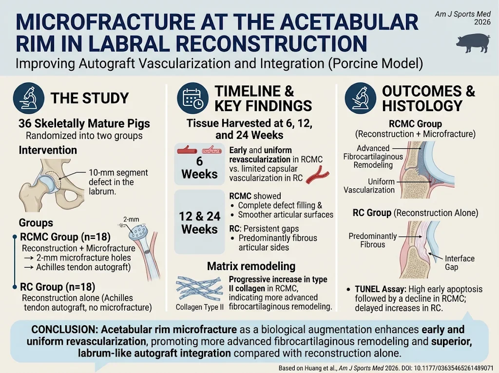
This controlled laboratory study utilized a randomized porcine model to investigate the effect of acetabular rim microfracture on labral autograft integration and vascularization. Thirty-six skeletally mature pigs were randomized into two groups: (1) Reconstruction with acetabular rim microfracture (RCMC), and (2) Reconstruction without microfracture (RC). A standardized 10-mm acetabular labral defect was created in each animal. In the RCMC group, the acetabular rim was microfractured by creating multiple 2-mm holes before reconstructing the labrum using autografts harvested from the Achilles tendon, secured at the rim. The RC group underwent identical reconstruction but without microfracture intervention. Specimens were harvested at 6, 12, and 24 weeks postoperatively for macroscopic and histological analyses focused on vascularization patterns, tissue remodeling, collagen composition, and apoptosis.Key Findings
Macroscopic observations revealed that at 12 and 24 weeks, the RCMC group demonstrated more complete defect filling and smoother articular surfaces compared to the RC group, indicating superior graft incorporation. Histological analysis showed that RCMC grafts underwent advanced fibrocartilaginous remodeling, featuring increased type II collagen—a marker of mature fibrocartilage—while RC grafts remained largely fibrous on the articular side with persistent gaps from native cartilage. Vascular distribution was notably different between groups. At 6 weeks, RCMC specimens showed uniform vascular infiltration throughout the graft tissue, whereas the RC group exhibited vascularization predominantly limited to the capsular side. By 24 weeks, both groups approached a native labrum-like peripheral vascular pattern; however, RCMC grafts achieved this state earlier and more uniformly. Apoptotic activity, assessed by terminal deoxynucleotidyl transferase dUTP nick-end labeling staining, was initially higher in the RCMC group, which may reflect active tissue remodeling and cellular turnover. Apoptosis gradually declined over time in RCMC samples, whereas the RC group exhibited a delayed increase in apoptosis, potentially signifying less favorable integration dynamics.Expert Commentary
The study provides compelling preclinical evidence that microfracture at the acetabular rim acts as a biological adjunct facilitating early vascular ingrowth and promoting fibrocartilaginous maturation of labral autografts. Microfracture likely enables access to bone marrow-derived progenitor cells and growth factors critical for tissue regeneration. Enhanced type II collagen deposition suggests that microfracture may shift healing from fibrous scar tissue toward more functional fibrocartilage, essential for labral biomechanical integrity. Limitations include the porcine model which, although relevant anatomically and physiologically, may not fully replicate human hip joint biomechanics or immune responses. Additionally, biomechanical data assessing graft strength and clinical studies evaluating functional outcomes are needed to corroborate these promising histological results. Future research should also investigate optimal microfracture techniques and timing, as well as potential adjunct therapies to maximize healing.Conclusion
Acetabular rim microfracture significantly enhances vascularization and integration of labral autografts in a porcine model, promoting more advanced fibrocartilaginous remodeling and labrum-like histological features than reconstruction alone. These findings support the potential of rim microfracture as a biological augmentation technique to improve labral reconstruction outcomes. However, translation to clinical practice requires further biomechanical validation and human trials to establish safety, efficacy, and functional benefits.Funding and Clinical Trial Registration
The study did not provide specific information about funding sources or clinical trial registration numbers.References
1. Jun H, Yiling T, Yonglie Z, et al. Microfracture at the Acetabular Rim as Biological Augmentation of Labral Reconstruction Improves Autograft Vascularization and Integration: A Porcine Model. Am J Sports Med. 2026 Nov 5:3635465261489071. doi: 10.1177/03635465261489071. PMID: 42989881. 2. Domb BG, Linder D, Jackson TJ. Update on Hip Labral Reconstruction: Indications, Techniques, and Outcomes. J Am Acad Orthop Surg. 2018;26(3):e50-e61. 3. Abrams GD, Frank RM, Gupta AK, et al. Arthroscopic management of labral tears and femoroacetabular impingement: Current concepts. J Am Acad Orthop Surg. 2015;23(12):706-717. 4. Steadman JR, Rodkey WG, Briggs KK. Microfracture: Surgical technique and rehabilitation to treat chondral defects. Clin Orthop Relat Res. 2001;(391 Suppl):S362-S369. 5. Philippon MJ, Schenker ML, Briggs KK, et al. Outcomes following arthroscopic labral reconstruction of the hip. Arthroscopy. 2014;30(5):646-650.Specialty calculators
For clinical reference; use professional judgment.