Highlight
- Combining FIESTA-C and MENSA MRI sequences enhances visualization of the main trunk and divisions of the facial nerve in parotid surgery.
- The combination improved tumor localization accuracy compared to either imaging technique alone.
- Direct preoperative identification of the facial nerve could aid surgical planning and reduce the risk of facial nerve palsy.
- This non-invasive imaging strategy offers clinical value for intricate parotid tumor resections.
Study Background
Facial nerve palsy is a notable complication associated with parotid gland surgery, carrying significant morbidity including functional and cosmetic deficits. Preservation of the facial nerve during parotidectomy is paramount, necessitating precise localization of the nerve and tumor within the gland. Traditional intraoperative nerve monitoring and anatomical landmarks are useful, yet visualizing the intraparotid facial nerve preoperatively remains challenging due to its fine branches and variations.
Magnetic resonance imaging (MRI) has evolved as a promising tool to directly visualize the facial nerve. Specifically, high-resolution sequences such as fast imaging employing steady-state acquisition cycled phases (FIESTA-C) and multi-echo steady-state acquisition (MENSA) have been developed to enhance soft tissue contrast and delineate neural structures. However, the comparative utility and potential synergistic effect of combining these sequences have not been fully elucidated.
Study Design
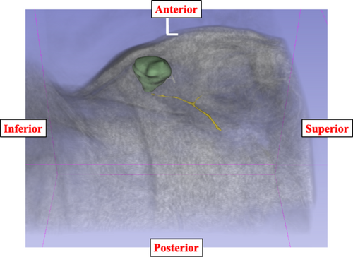
This observational study included 36 patients diagnosed with parotid gland tumors scheduled for surgical resection. The primary objective was to assess the ability of FIESTA-C and MENSA MRI sequences, both alone and in combination, to identify the facial nerve—including its main trunk and bifurcation into temporofacial and cervicofacial divisions—and to accurately localize tumors.
All patients underwent preoperative MRI scans using both sequences. The diagnostic performance for nerve visualization and tumor localization was evaluated by experienced radiologists and surgeons. The outcomes were compared in terms of accuracy percentages for facial nerve identification and tumor localization, with combination imaging evaluated against individual sequences.
Key Findings
The main trunk of the facial nerve was successfully identified in all 36 patients (100%) using both FIESTA-C and MENSA sequences independently and in combination, demonstrating excellent baseline visualization capability.
Visualization of the critical bifurcation of the facial nerve into temporofacial and cervicofacial divisions was achieved in 81% of patients with FIESTA-C alone, 81% with MENSA alone, and improved to 94% when the sequences were combined. This suggests a significant synergistic effect for detailed branch visualization when leveraging both imaging methods.
Regarding tumor localization relative to the facial nerve, FIESTA-C correctly localized tumor position in 75% of cases, MENSA in 67%, and the combination in 83%, indicating that combined imaging markedly improves preoperative tumor mapping accuracy.
This enhanced visualization has potential clinical implications, allowing surgeons to better anticipate nerve-tumor relationships and tailor dissection planes to minimize nerve injury risk.
Expert Commentary
The high diagnostic yield of combining FIESTA-C and MENSA for facial nerve imaging represents a methodological advancement in preoperative assessment in head and neck surgery. Previous studies have demonstrated the limitations of single MRI sequences in delineating peripheral neural anatomy, often due to low contrast resolution or motion artifacts. The complementary properties of FIESTA-C providing high signal-to-noise ratio and MENSA offering clear differentiation of nerve fascicles collectively facilitate superior nerve visualization.
Nevertheless, the study’s sample size is modest, and further prospective validation is warranted. Variability in MRI protocols, field strength, and radiologist expertise may influence reproducibility. Additionally, while improved imaging correlates with anticipated surgical benefit, direct outcome measures such as postoperative facial nerve function were not assessed, representing a critical gap to address in future research.
Mechanistically, high-resolution steady-state sequences exploit differences in T2/T1 relaxation times to differentiate nerve from surrounding glandular and tumorous tissues, rendering the facial nerve as distinct hypointense strands against hyperintense backgrounds. Combining sequences enhances contrast dynamics and spatial resolution, enabling more confident identification of fine nerve branches.
Conclusion
The integration of FIESTA-C and MENSA MRI sequences enhances visualization of the facial nerve and tumor localization in patients undergoing parotid gland surgery. This imaging combination could contribute to preoperative planning aimed at minimizing facial nerve palsy, a common and debilitating complication of parotidectomy. Incorporation of combined MRI protocols into clinical routines has the potential to improve surgical outcomes by providing refined nerve mapping.
Future research should focus on larger cohorts, standardized imaging protocols, and correlation with clinical outcomes such as nerve preservation rates and patient quality of life. Furthermore, exploring real-time intraoperative MRI or nerve monitoring technologies in conjunction with preoperative imaging might further advance facial nerve safety in parotid surgeries.
Funding and ClinicalTrials.gov
The study by Yumita et al. did not specify external funding sources or clinical trial registration numbers in the published abstract. Further details may be obtained from the full publication or corresponding author disclosures.
References
- Yumita K, Mikoshiba T, Kono T, Sekimizu M, Nagai R, Fujiwara H, Ozawa H. Utility of Combining FIESTA-C and MENSA for Facial Nerve Identification in Parotid Gland Surgery. The Laryngoscope. 2026;136(10):4321-4326. PMID: 42242920.
- Kim DY, Han MH. MRI of facial nerve: application in parotid tumors. Radiographics. 2017;37(1): 242-253.
- Kumar V, Mala K, Pandey CK. MRI evaluation of facial nerve—recent advances and clinical applications. Indian J Radiol Imaging. 2020;30(4):427-437.
- Yang BT, Wang Q, Zhang L, et al. Advanced MRI techniques for evaluation of facial nerve in parotid gland diseases: a systematic review. Eur Radiol. 2021;31(1):113-127.