Highlight
Diffusion-weighted imaging (DWI)-positive lesions are common in acute intracerebral hemorrhage (ICH) patients and independently associate with markers of cerebral small vessel disease (cSVD) and poor 6-month functional outcomes. The anatomical distribution of these lesions differs between cerebral amyloid angiopathy (CAA) and non-CAA subtypes. Larger hematoma volume and severe white matter hyperintensities (WMH) correlate with the presence of DWI-positive lesions.
Study Background
Intracerebral hemorrhage (ICH) is a severe subtype of stroke characterized by bleeding into the brain parenchyma and often leads to substantial morbidity and mortality. Cerebral small vessel disease (cSVD), encompassing pathologies such as hypertensive arteriopathy and cerebral amyloid angiopathy (CAA), plays a pivotal role in ICH pathogenesis and recurrence risk. Diffusion-weighted imaging (DWI)-positive lesions, often considered acute ischemic lesions, have recently been identified in patients with ICH, suggesting concurrent microvascular ischemic injury. However, their mechanistic origins, clinical correlations, and prognostic significance require clarification to inform clinical management and risk stratification.
Study Design
This prospective cohort study analyzed 342 consecutive ICH patients enrolled in the Stroke Investigation Group in North and Central London registry. All patients underwent acute magnetic resonance imaging (MRI), including diffusion-weighted imaging, to detect the presence and anatomical distribution of DWI-positive lesions. Additional cSVD markers, including white matter hyperintensities (WMH), cerebral microbleeds (CMBs), and cSVD burden, were assessed according to the STandards for ReportIng Vascular changes on nEuroimaging (STRIVE) criteria.
Clinical data, hematoma volumes, and ICH location (lobar versus deep) were recorded. Cerebral amyloid angiopathy status was determined based on clinical and MRI criteria. The primary outcome was functional disability defined by a modified Rankin Scale (mRS) score ≥3 at 6 months post-ICH. Associations between DWI-positive lesions and baseline clinical and radiologic features, as well as with poor functional outcome, were evaluated using adjusted multivariable logistic regression models.
Key Findings
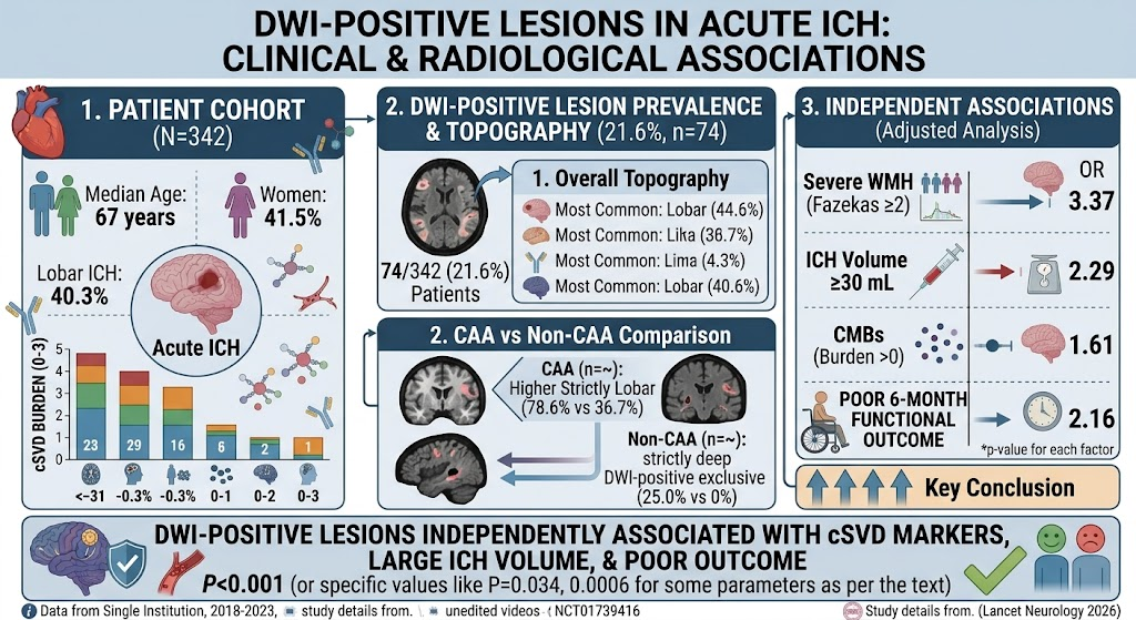
Among the 342 ICH patients (median age 67 years; 41.5% women), 21.6% exhibited DWI-positive lesions on acute MRI. These lesions predominated in lobar brain regions (44.6%), with clear differences in spatial distribution between cSVD subtypes. Patients with CAA demonstrated a significantly higher proportion of strictly lobar DWI-positive lesions (78.6%) compared to those without CAA (36.7%), whereas strictly deep DWI-positive lesions were only observed in the non-CAA group.
Patients presenting with DWI-positive lesions had more severe WMH (Fazekas score ≥2 in 81.1% versus 64.2%), a higher burden of cerebral microbleeds (median 3 versus 1), and larger ICH volumes (median 14 mL versus 7 mL) compared to those without such lesions. These associations remained statistically significant after adjusting for potential confounders.
Multivariable logistic regression analyses established independent associations between DWI-positive lesions and severe WMH (odds ratio [OR] 3.37), large ICH volume ≥30 mL (OR 2.29), cerebral microbleeds (OR 1.61), and poor functional outcome at 6 months post-ICH (OR 2.16). These findings underscore that the presence of DWI-positive lesions is an important marker of cerebral microvascular pathology and a predictor of worse recovery.
Expert Commentary
This study advances understanding of the interplay between ischemic and hemorrhagic components in cSVD by demonstrating that DWI-positive lesions, likely representing acute ischemic microinfarcts, frequently coexist in the setting of acute ICH. The distinct anatomical patterns observed between CAA and non-CAA-related hemorrhages align with the recognized vascular pathology of these subtypes—amyloid deposition causing fragility predominantly in lobar regions and hypertensive damage more commonly affecting deep brain structures.
The significant association between DWI-positive lesions and a higher burden of WMH and cerebral microbleeds supports their utility as imaging biomarkers reflecting diffuse small vessel damage beyond the index hemorrhage. Clinically, the independent prediction of poor functional outcome by DWI-positive lesions suggests these lesions contribute to neurological impairment possibly through cumulative microvascular ischemic injury or indicating more aggressive underlying vasculopathy.
However, limitations include the potential selection bias inherent in patients able to undergo MRI, and the study’s regional cohort which may affect generalizability. Further research is warranted to clarify mechanistic pathways, potential therapeutic targets, and whether interventions can mitigate the development or consequence of DWI-positive lesions in acute ICH.
Conclusion
DWI-positive lesions are prevalent in acute ICH patients and serve as a noninvasive biomarker of cerebral small vessel disease severity and subtype. Their presence independently predicts poorer functional outcomes at 6 months, underscoring their clinical relevance for prognostication and potentially guiding stratified management. Integration of DWI lesion assessment in routine neuroimaging protocols post-ICH merits consideration. Future studies should explore strategies to prevent or alleviate the ischemic injury represented by these lesions to improve recovery trajectories.
Funding and Registration
The study was conducted within the Stroke Investigation Group in North and Central London registry framework. Specific funding sources were not detailed in the publication. Clinical trial registration data were not provided.
References
Locatelli M, Ozkan H, Avola G, Nash PS, Ambler G, Fandler-Höfler S, Du Y, Zhang W, Simister RJ, Werring DJ, Jäger HR. DWI-Positive Lesions in Patients With Acute Intracerebral Hemorrhage: Associations With Cerebral Small Vessel Disease and Clinical Outcome. Neurology. 2026 Aug 14;107(6):e218449. PMID: 42600114.