Neurotransmitter-Guided Connectome Disruption as a Key to Poststroke Language Impairment

Highlight

  • Poststroke language outcomes vary widely and are inadequately explained by lesion location alone.
  • Structural network disruption informed by neurotransmitter receptor distribution revealed key roles of serotonergic (5-HT1a, 5-HT2a) and dopaminergic (D1) systems in language impairment.
  • Neurotransmitter-driven connectome damage explained additional variability in language deficits beyond traditional clinical factors.
  • Findings support a novel neurochemical network framework for understanding poststroke aphasia with potential, though preliminary, implications for targeted interventions.

Study Background

Stroke remains a leading cause of adult disability, with aphasia affecting up to one-third of survivors, severely impacting communication and quality of life. Despite advances in neuroimaging and clinical assessment, predicting the extent and recovery trajectory of language impairment remains challenging. Conventional approaches predominantly focus on lesion size and cortical location within classical language areas, yet significant unexplained variability persists. Emerging evidence suggests that beyond focal lesions, disruption to distributed brain networks—especially those involving neurochemical systems integral to synaptic transmission and plasticity—may critically influence language outcomes. Understanding the neurochemical underpinnings and structural connectivity profiles related to these systems after stroke may enhance prognostication and inform rehabilitation strategies.

Study Design

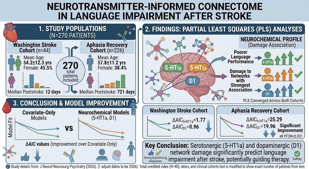
This study analyzed two distinct cohorts of patients post left-hemispheric stroke, encompassing acute and chronic stages of recovery. The Washington Stroke Cohort (n=44) included patients assessed around 1-2 weeks post first symptomatic stroke using a comprehensive language battery. The Aphasia Recovery Cohort (n=226) involved patients in the chronic phase (median ~2 years poststroke), evaluated with the Western Aphasia Battery-Revised. Individual lesion masks were embedded into normative structural connectomes weighted by positron-emission tomography (PET)-derived density maps of sixteen neurotransmitter receptors and transporters, including serotonergic and dopaminergic markers. Partial least squares regression and multivariable linear models (adjusted for age, sex, lesion volume, and time poststroke) were used to identify neurotransmitter-informed network disruptions associated with language performance.

Key Findings

A total of 270 patients were included with similar demographic profiles across cohorts. Partial least squares analyses consistently highlighted neurochemical profiles involving serotonergic receptors (notably 5-HT1a and 5-HT2a) and dopaminergic D1 receptors as the principal predictors of poststroke language impairment. These neurotransmitter-related network disruptions showed statistically significant associations with poorer language scores in both acute (Washington Stroke Cohort) and chronic (Aphasia Recovery Cohort) stages.

In fully adjusted regression models, damage to 5-HT1a and D1 receptor-weighted networks remained significant despite controlling for key clinical variables and lesion burden, improving model fits considerably (all false discovery rate-corrected P values <0.001). The improvement in Akaike information criterion (AIC) indicated better explanatory power of models incorporating neurotransmitter-informed network damage compared to classical predictors alone (eg, ∆AIC for 5-HT1a was 1.77 in acute and 25.29 in chronic cohorts).

These findings suggest that poststroke language deficits are not solely attributable to lesion location or size but also to the disruption of specific neurochemical pathways, particularly serotonergic and dopaminergic systems. This adds a novel dimension to the connectome perspective by integrating neurotransmitter maps derived from PET imaging, effectively providing a structural proxy for neurochemical integrity.

Expert Commentary

These novel insights into the neurochemical underpinnings of poststroke aphasia have important implications. The serotonergic and dopaminergic systems modulate synaptic plasticity, cognitive processing, and motivation—functions critical for language rehabilitation. The demonstration that damage to 5-HT1a and D1 receptor-related networks predicts language outcomes suggests that pharmacological modulation of these neurotransmitter systems might enhance recovery when combined with speech therapy.

However, the study’s indirect use of normative PET maps as proxies for neurotransmitter distribution in individual patients is an important limitation, as actual receptor densities may vary significantly due to age, stroke-induced changes, or medication effects. Additionally, the observational design precludes inference of causality or therapeutic efficacy. Future studies incorporating in vivo PET imaging poststroke alongside longitudinal language assessments could validate and refine these associations.

Conclusion

This study provides compelling evidence that disruption of serotonergic (especially 5-HT1a) and dopaminergic (D1) neurotransmitter-informed networks contributes significantly to language impairment after stroke, beyond traditional clinical and lesion metrics. Integrating neurochemical connectomics with clinical neuroimaging may enhance prediction accuracy and guide novel rehabilitation approaches targeting these neurotransmitter systems. Despite these advances, further research is necessary to translate these findings into validated clinical interventions.

Funding and Registration

The study analyzed openly available datasets including the Washington Stroke Cohort and Aphasia Recovery Cohort. Detailed funding sources and clinical trial registrations were not reported in the primary publication.

References

  • Hornberger T, Schulz R, Koch PJ, et al. Neurotransmitter-Informed Connectome Approach to Language Impairment After Stroke. Stroke. 2026;57(8):2482-2492. PMID: 42283088.
  • Berthier ML. Poststroke aphasia: epidemiology, pathophysiology and treatment. Drugs Aging. 2005;22(2):163-182.
  • Crosson B, McGregor K, Gopinath K, et al. Functional Imaging and Frontotemporal Dementia: Implications for Rehabilitation. J Head Trauma Rehabil. 2014;29(5):E23-32.
  • Rossini PM, Burke D, Chen R, et al. Non-invasive electrical and magnetic stimulation of the brain, spinal cord, roots and peripheral nerves: basic principles and procedures for routine clinical and research application. Clin Neurophysiol. 2015;126(6):1071-1107.
  • Maurer K, Marten-Mittag B, Stieltjes B, et al. A PET and MRI study of poststroke aphasia recovery: functional and structural changes in language networks. Brain Lang. 2019;194:45-54.

Comments

No comments yet. Why don’t you start the discussion?

Leave a Reply