Highlight
This retrospective multicenter case series identified a previously undescribed ultrasound pattern—discontinuous peripheral punctate echogenic foci (DPEF)—in thyroid nodules. All nodules exhibiting this pattern were confirmed histopathologically as papillary thyroid carcinoma (PTC). This novel sonographic feature may enhance malignancy risk stratification and warrants broader recognition among thyroid imaging specialists.
Study Background
Thyroid nodules are common clinical findings, and ultrasonography (US) remains the primary imaging modality to stratify malignancy risk. Current ultrasound risk stratification systems describe various types of echogenic foci such as microcalcifications (punctate echogenic foci within the nodule), coarse calcifications, and complete or incomplete peripheral (rim) calcifications. While these characteristics help guide clinical management, ultrasonography is limited by imperfect sensitivity and specificity. The discovery of new sonographic patterns that can reliably indicate malignancy could improve diagnostic accuracy and reduce unnecessary interventions.
Papillary thyroid carcinoma (PTC) is the most common thyroid malignancy and often demonstrates microcalcifications on ultrasound, which are considered a hallmark feature. However, peripheral calcifications traditionally associate poorly with malignancy, limiting their utility in risk assessments. The identification of discontinuous peripheral punctate echogenic foci (DPEF) introduces a potentially important sonographic pattern correlating with PTC, challenging prior assumptions about peripheral calcifications.
Study Design
This multicenter retrospective case series collated thyroid ultrasound images from five international centers across Brazil, United States, Italy, France, and Romania from January 2020 through September 2025. Inclusion criteria required the presence of discontinuous peripheral punctate hyperechoic foci—small, discrete echogenic dots arrayed discontinuously along the nodule periphery—irrespective of nodule echogenicity or other morphological features.
Each thyroid nodule underwent multidisciplinary review, including expert radiologists, endocrinologists, head and neck surgeons, and pathologists, to ensure comprehensive interpretation. Final diagnoses were confirmed through histopathological analysis of surgically resected specimens, providing a robust gold standard reference for malignancy status.
Key Findings
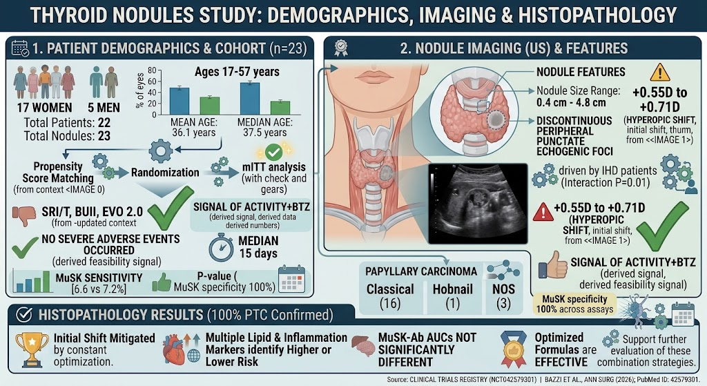
A total of 23 nodules from 22 patients (17 females, 5 males; mean age 36.1 years) were included. Nodule sizes ranged from 0.4 cm to 4.8 cm. On high-resolution ultrasound, all nodules exhibited the distinctive pattern of small, multiple echogenic foci arranged discontinuously along their peripheral margins, differing from classical patterns of microcalcifications or complete rim calcifications.
Histopathology confirmed that all nodules (100%) were papillary thyroid carcinomas. Subtypes identified included 16 classical PTC variants, one hobnail variant, and three PTCs not otherwise specified. There was no evidence of benign pathology or other malignancies in association with DPEF. This finding points to a strong and specific correlation between DPEF and PTC presence.
The study did not directly compare sensitivity or specificity metrics against conventional ultrasound features, as its design was descriptive and hypothesis-generating. Nonetheless, the uniform presence of PTC in all nodules exhibiting DPEF signals a potentially high positive predictive value.
Expert Commentary
The identification of DPEF represents a significant refinement in thyroid ultrasound assessment. Conventional risk stratification tools emphasize intranodular microcalcifications as a hallmark of PTC but generally consider peripheral calcifications less suspicious. This study challenges that view by demonstrating that a previously unrecognized pattern—small, discontinuous echogenic spots along the nodule edge—may be a specific indicator of malignancy.
From a pathophysiological standpoint, these peripheral echogenic foci could represent early or fragmented calcifications or psammoma bodies situated along the nodule boundary. Molecular and histological correlation studies would be valuable to elucidate the biological basis of DPEF and validate its diagnostic utility.
Limitations include the retrospective nature, small sample size, potential selection bias, and lack of comparative data with other benign nodules featuring peripheral calcifications. Future larger prospective studies should evaluate diagnostic accuracy parameters of DPEF compared with established features and test reproducibility across diverse populations and ultrasound equipment.
Conclusion
This multinational retrospective case series describes a novel ultrasound pattern—discontinuous peripheral punctate echogenic foci—that is strongly associated with papillary thyroid carcinoma. Recognition of DPEF may improve risk stratification and clinical decision-making in thyroid nodule evaluation, pending validation by future studies quantifying its sensitivity and specificity.
Reporting this pattern may raise awareness among sonographers and radiologists, facilitating earlier identification of PTC and potentially reducing unnecessary biopsies or surgeries in patients without this feature. Ultimately, integrating DPEF into multimodal imaging algorithms could refine thyroid cancer diagnosis and optimize patient management.
Funding and ClinicalTrials.gov
The study was a collaborative effort across multiple academic and clinical centers. Specific funding sources or clinical trial registrations were not reported in the manuscript.
References
1. Daibes L, Spiezia S, Nascimento MF, et al. Discontinuous Peripheral Punctate Echogenic Foci in Thyroid Nodules: A New Sonographic Pattern Suggestive of Papillary Carcinoma. Thyroid. 2026 Aug 13:10507256261477161. PMID: 42592648.
2. Tessler FN, Middleton WD, Grant EG, et al. ACR Thyroid Imaging, Reporting and Data System (TI-RADS): White Paper of the American College of Radiology. J Am Coll Radiol. 2017;14(5):587-595.
3. Haugen BR, Alexander EK, Bible KC, et al. 2015 American Thyroid Association Management Guidelines for Adult Patients with Thyroid Nodules and Differentiated Thyroid Cancer. Thyroid. 2016;26(1):1-133.