Highlight
– Upper cervical cord area (MUCCA) decline in multiple sclerosis (MS) exceeds normal aging effects, especially notable in the fifth and sixth decades.
– Disease-specific atrophy patterns are independent of sex and largely influenced by disease duration rather than age at onset.
– Early-onset MS patients show lower normalized MUCCA Z-scores compared to adult and late-onset, but differences diminish after adjusting for disease duration.
– MUCCA atrophy strongly correlates with clinical disability and structural MRI abnormalities, supporting its role as a sensitive biomarker of MS progression beyond aging.
Study Background
Multiple sclerosis (MS) is a chronic neuroinflammatory and neurodegenerative disorder characterized by demyelination and axonal loss within the central nervous system (CNS). Spinal cord involvement is common and contributes significantly to disability. Cervical cord atrophy, particularly at the upper cervical levels (C1-C3), is a known predictor of disability progression and motor impairment in MS patients. However, the extent to which this atrophy is driven by normal aging versus disease-specific neurodegeneration has been unclear. Age-related spinal cord changes could confound interpretation of MRI-based cord measurements, limiting their utility in clinical trials and practice. Furthermore, heterogeneity related to sex differences and age at MS onset complicates assessment of cervical cord atrophy patterns. This study aimed to disentangle age-related from disease-specific upper cervical cord atrophy in MS, exploring trajectory changes with aging, sex, onset age categories, and clinical correlates, to refine the role of upper cervical cord measures as biomarkers in MS management.
Study Design
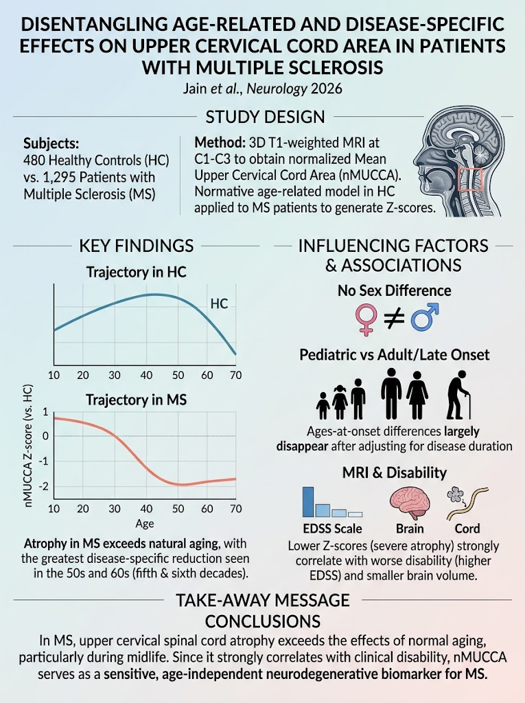
This was a cross-sectional, multicenter study conducted across three Italian sites. The study included 1,295 patients with clinically confirmed MS and 480 healthy controls (HCs), aged 18-70 years, all free from other spinal cord pathologies. High-resolution 3.0 Tesla T1-weighted brain MRIs were used to derive volumetric measurements of the mean upper cervical cord area (MUCCA) between C1 and C2/3 vertebral levels. MUCCA values were normalized for head size (nMUCCA). A polynomial regression model was fitted on HC data to characterize normative lifespan trajectories of nMUCCA, adjusting for age, age squared, sex, scanner variability, and interaction terms. This model generated Z-scores for MS patients, reflecting deviation from expected age- and sex-matched norms. MS patients were categorized by age at disease onset into pediatric onset (<18 years), adult onset (18–49 years), and late onset (≥50 years). Associations between nMUCCA Z-scores and clinical disability, disease duration, T2 lesion burden, as well as normalized brain volumes were analyzed.
Key Findings
Among healthy controls, nMUCCA exhibited a nonlinear trajectory with age: an increase up to the late 30s followed by a decline starting after 50 years. This pattern confirms that cervical cord area is subject to physiological growth during young adulthood and involution with aging. In MS patients, nMUCCA Z-scores fell significantly below zero from age 30 years onward, indicating disease-specific cord atrophy beyond normal aging. The most pronounced reductions were observed in the fifth and sixth decades of life. The disease-associated decline rate was steepest in early adulthood extending to the late 40s, with slowing attenuation thereafter likely due to overlapping aging effects.
Sex did not significantly modify these patterns, suggesting no sex-specific differences in cord atrophy trajectories among MS patients. Pediatric-onset MS patients had the lowest nMUCCA Z-scores compared to adult- and late-onset groups, but this difference became nonsignificant after adjusting for disease duration, implicating longer disease exposure rather than intrinsic onset age effects.
Correlation analyses revealed that more severe upper cervical cord atrophy is significantly associated with higher disability scores, longer disease duration, greater T2 lesion load, and lower normalized brain volumes (correlation coefficients ranging from 0.196 to 0.392, all statistically significant with p-values < 0.001). These findings underscore the clinical and structural relevance of cervical cord atrophy as a surrogate marker of neurodegenerative burden in MS.
Expert Commentary
This study elegantly addresses a critical confounder in spinal cord imaging for MS — the physiologic influence of aging. By employing robust statistical modeling of healthy control data and applying Z-score metrics, the authors successfully isolate the disease-specific atrophic component. Understanding that upper cervical cord atrophy in MS patients exceeds expected aging-related shrinkage advances the biomarker validation necessary for clinical application.
The absence of sex effects on cord atrophy trajectory contrasts with some previous neuroimaging studies reporting sex differences in brain atrophy patterns, suggesting spinal cord involvement may be more uniformly impacted across sexes in MS. The finding that pediatric onset patients demonstrate greater cord atrophy primarily reflects longer disease duration, emphasizing the cumulative nature of neurodegeneration rather than developmental vulnerability.
The strong correlation of MUCCA measures with established MRI and disability markers supports their translational utility for monitoring disease progression and potentially evaluating neuroprotective interventions targeting spinal cord pathology. However, the cross-sectional design limits direct causal inference and tracking of individualized atrophy progression over time. Further longitudinal studies are warranted to confirm trajectories and temporal dynamics. Additionally, the study population was drawn from Italian centers; replication in diverse ethnic and racial cohorts would enhance generalizability.
Conclusion
This comprehensive study establishes that upper cervical cord atrophy in MS patients is partially independent from normal aging and strongly correlates with clinical disability and CNS structural damage. The nonlinear age trajectories and prominent atrophy during mid-life provide insights into critical windows for potential therapeutic intervention. Disease-specific cord atrophy is not influenced by sex and differences across age-at-onset groups are largely explained by disease duration rather than intrinsic onset age effects. These findings reinforce upper cervical cord area as a sensitive and clinically meaningful biomarker of neurodegeneration in MS, with potential applications in routine care and clinical trials.
Funding and Trial Registration
Details regarding the study’s funding sources and clinical trial registration were not specified in the source article.
References
Jain K, Storelli L, Valsasina P, Preziosa P, Meani A, Pantano P, Piervincenzi C, Gallo A, D’Ambrosio A, Filippi M, Rocca MA. Disentangling Age-Related and Disease-Specific Effects on Upper Cervical Cord Area in Patients With Multiple Sclerosis. Neurology. 2026 Sep 30;107(8):e218517. doi:10.1212/WNL.0000000000018517. PMID: 42814910.