
Introduction
Systemic amyloidoses represent a heterogeneous group of protein misfolding disorders characterized by extracellular deposition of insoluble amyloid fibrils in various organs. Among these, light chain (AL) amyloidosis is the most common form related to monoclonal immunoglobulin light chains (LCs) secreted by clonal plasma cells. Early and precise detection of amyloidogenic LCs is paramount because AL amyloidosis, although rapidly progressive, is amenable to effective treatments that can preserve organ function and improve survival. This review synthesizes advances in the detection, monitoring, and discrimination between amyloidogenic and benign (“innocent”) light chains in systemic amyloidoses, highlighting clinical implications and future prospects.
Clinical Context and Disease Burden
Monoclonal gammopathies span a clinical spectrum, from benign monoclonal gammopathy of undetermined significance (MGUS) to malignant conditions such as multiple myeloma and Waldenström macroglobulinemia. In some patients, pathogenic monoclonal immunoglobulins induce organ damage—a clinical entity termed monoclonal gammopathies of clinical significance (MGCS). AL amyloidosis arises when a clone produces amyloidogenic LCs prone to misfolding and fibril formation, leading to multi-organ dysfunction, commonly involving the heart, kidneys, liver, and peripheral nerves.
The prevalence of AL amyloidosis is low relative to other plasma cell disorders but its impact is profound due to rapid progression and poor prognosis if untreated. Cardiac involvement is the major determinant of mortality. Early diagnosis remains challenging due to nonspecific symptoms and the need for sensitive and specific laboratory detection tools.
Laboratory Detection of Amyloidogenic Light Chains
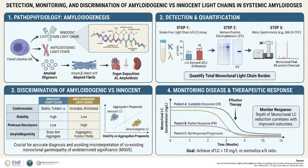
Diagnosis depends on the demonstration of a monoclonal LC that forms amyloid deposits in tissues. Traditional methods include immunofixation electrophoresis and serum free light chain (FLC) assays. While these are essential screening tools, their sensitivity and specificity can be limited, especially in cases with low clonal burden or renal impairment affecting FLC metabolism.
Recent advances in mass spectrometry (MS)-based proteomics have transformed LC detection. MS provides high sensitivity and specificity, enabling identification of clonal LCs directly from serum or tissue biopsies and confirming amyloidogenic LC presence. Flow cytometry and next-generation sequencing (NGS) approaches facilitate deep hematologic response assessment by detecting minimal residual disease (MRD), guiding therapy cessation, or modification.
Monitoring Treatment Response and Relapse
Quantitative assays of LC levels are vital to monitor hematologic response to therapy, detect relapse, and assess measurable residual disease. The therapeutic landscape has rapidly evolved with proteasome inhibitors, immunomodulatory drugs, and monoclonal antibodies achieving deeper hematologic responses. Sensitive methods detecting low-level residual clonal LCs improve prognostic accuracy and enable personalized management.
Despite these advances, relapse definitions remain non-standardized, representing an unmet clinical need. Standardization across assays and incorporation of MRD techniques into routine practice may optimize patient outcomes.
Discriminating Amyloidogenic Versus Innocent Light Chains
A major clinical challenge lies in differentiating benign LCs—common in MGUS and other plasma cell disorders—from those prone to form amyloid fibrils. Identification of patients at risk before clinical organ involvement allows for pre-symptomatic intervention, potentially altering disease trajectory.
Emerging discriminators include biomarkers of early organ damage (e.g., NT-proBNP for cardiac strain, albuminuria for renal involvement) and intrinsic LC characteristics:
- N-linked glycosylation: Amyloidogenic LCs frequently exhibit aberrant glycosylation patterns influencing folding and aggregation propensity.
- Germline gene usage: Certain VH and VL gene families are preferentially associated with amyloidogenic clones.
- Structural stability: Reduced thermodynamic stability correlates with increased amyloidogenic potential.
Application of these biomarkers combined with clinical risk stratification could facilitate early diagnosis and tailored treatment.
Differential Diagnosis and Clinical Implications
Transthyretin (ATTR) cardiac amyloidosis is prevalent in elderly males and African-American populations and often coexists with monoclonal gammopathies producing LCs. Distinguishing AL from ATTR amyloidosis is critical since treatment strategies diverge substantially.
Laboratory approaches integrating LC detection with molecular amyloid typing enable rapid exclusion or confirmation of AL amyloidosis. This clinical imperative minimizes inappropriate chemotherapy administration in ATTR patients and expedites AL-directed therapy in true light chain amyloidosis.
Expert Commentary and Future Directions
Recent advances enhance hematologic diagnostic sensitivity and specificity, yet challenges remain in standardizing relapse definitions and deploying predictive biomarkers broadly. Integrating mass spectrometry with immunophenotyping and molecular profiling offers a comprehensive toolbox for clinicians.
Further research into LC-intrinsic features and their relationship to amyloid formation may unlock therapeutic targets to prevent amyloidogenesis at the molecular level. Additionally, longitudinal studies assessing biomarker-driven preemptive therapies in MGUS patients at risk for AL amyloidosis are warranted.
Conclusions
Effective management of systemic amyloidoses requires precise laboratory tools to detect, quantify, and distinguish amyloidogenic LCs. Advances in mass spectrometry, flow cytometry, and next-generation sequencing are reshaping diagnostic and monitoring paradigms. Discrimination between innocent and amyloidogenic light chains through biomarker and molecular profiling heralds a new era of early diagnosis and personalized interventions to improve organ preservation and survival in AL amyloidosis.
References
- Palladini G, Milani P, Nuvolone M. Detection, Monitoring, and Discrimination of Amyloidogenic vs Innocent Light Chains in Systemic Amyloidoses. Blood. 2026 Nov 9. PMID: 43002606.
- Kumar S, Dispenzieri A, Lacy MQ, et al. Clonal assessment in AL amyloidosis: disease monitoring and prognostic impact. Blood. 2021;137(1):57-65.
- Merlini G, Palladini G. Amyloidosis: Pathogenesis and New Therapeutic Options. J Clin Oncol. 2020;38(33):3886-3899.
- Sidana S, Kumar S. Monitoring response in AL amyloidosis: current and emerging strategies. Hematol Oncol Clin North Am. 2023;37(3):451-465.
Specialty calculators
For clinical reference; use professional judgment.