Highlight
- Myelodysplastic neoplasms (MDS) provoke profound spatial alterations in bone marrow microarchitecture beyond blast count abnormalities.
- Multiplex immunofluorescence combined with single-cell phenotyping enables quantification of >5 million spatially resolved marrow cells in patients and controls.
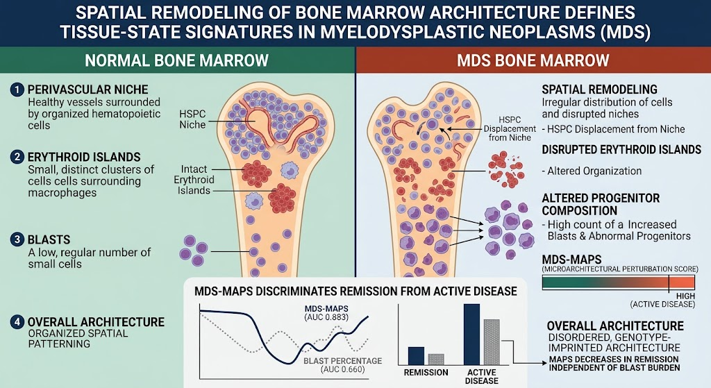
- A novel Microarchitectural Perturbation Score (MDS-MAPS) summarizes 82 architectural features, outperforming blast percentage in distinguishing active disease from remission and low-blast MDS from clonal cytopenia of undetermined significance.
- MDS-MAPS dynamically tracks marrow remodeling during treatment, correlating with disease activity independently of blast burden and indicating tissue-state normalization or relapse.
Study Background
Myelodysplastic neoplasms (MDS) represent a heterogeneous group of clonal hematopoietic disorders characterized by ineffective hematopoiesis, peripheral cytopenias, and a risk of progression to acute myeloid leukemia. Current clinical evaluation relies primarily on blast percentage assessment and qualitative morphological evaluation of bone marrow biopsies. However, such assessments incompletely reflect the complexity of marrow microenvironment alterations, which include disrupted cellular niches and aberrant spatial arrangements of hematopoietic progenitors that critically influence disease biology and therapeutic responses.
Modern imaging and analysis techniques have not routinely been integrated into clinical practice for MDS due to technical complexity and interpretive challenges. Yet, capturing spatial compositional and architectural changes could improve diagnostic accuracy, disease stratification, and real-time monitoring of treatment efficacy. This study addresses this unmet need by applying advanced multiplex immunofluorescence imaging combined with computational single-cell spatial phenotyping to characterize and quantify marrow architecture in MDS compared with precursor states and normal controls.
Study Design
This translational research study analyzed archival diagnostic and longitudinal bone marrow biopsy samples from patients with MDS, precursor conditions (clonal cytopenia of undetermined significance [CCUS]), and healthy controls. The total cohort included 36 diagnostic MDS samples, 29 longitudinal treatment follow-up samples, 13 precursor state samples, and 21 normal control biopsies. Over 5 million spatially resolved single cells were profiled using whole-slide multiplex immunofluorescence imaging that allowed simultaneous visualization of multiple cell markers.
The analytic workflow involved phenotype classification of hematopoietic populations, spatial mapping of cellular niches, and identification of microarchitectural features such as progenitor cell composition, erythroid island integrity, and localization of hematopoietic stem and progenitor cells (HSPCs) relative to vascular niches. An algorithmic Microarchitectural Perturbation Score (MDS-MAPS) was derived based on 82 quantitative spatial and cellular features extracted from baseline diagnostic samples. This score was then validated in longitudinal samples to assess correlation with clinical disease activity.
Key Findings and Results
The study identified significant, genotype-imprinted remodeling of bone marrow architecture in MDS compared to controls and precursor states. Key observations included:
– Altered Progenitor Composition and Spatial Distribution: MDS samples demonstrated a shift in progenitor cell populations with abnormal spatial clustering and depletion in typical stem cell vascular niches.
– Disrupted Erythroid Islands: The classic structural arrangement of erythroid precursors was markedly perturbed, reflecting ineffective erythropoiesis characteristic of MDS.
– Displacement of HSPCs: Hematopoietic stem and progenitor cells, normally enriched in perivascular niches, showed aberrant localization, indicating niche disruption.
The composite MDS-MAPS, integrating multiple cellular and spatial parameters, distinguished active MDS from remission states more accurately than conventional blast percentage metrics, with an area under the curve (AUC) of 0.883 versus 0.660. Furthermore, MDS-MAPS effectively separated low-blast MDS cases from CCUS with an AUC of 0.815, highlighting its potential for refined diagnostic discrimination.
Longitudinal analyses revealed that MDS-MAPS decreased significantly upon remission independent of blast count reductions, suggesting architectural normalization. Conversely, relapse was marked by re-emergence of architectural perturbations detected by increasing MAPS values. Mixed-effects modeling confirmed these findings, underscoring MDS-MAPS as a dynamic tissue-state biomarker reflecting disease status beyond traditional morphological assessment.
Expert Commentary
This study advances our understanding of how spatial bone marrow architecture can serve as a biomarker for MDS disease activity and therapeutic response. By integrating high-dimensional imaging with quantitative spatial analysis, the authors move beyond static blast counts toward a biologically nuanced metric capturing microenvironmental alterations central to MDS pathogenesis.
However, translating such complex imaging techniques into routine clinical workflows remains a challenge due to technical demands and the need for standardization. Larger prospective studies are warranted to validate MDS-MAPS across diverse patient populations and treatment regimens. Additionally, mechanistic insights linking specific architectural changes to molecular pathways or clinical outcomes would strengthen the biological plausibility and potential clinical utility of these findings.
Despite these limitations, the methodology exemplifies how spatial tissue phenotyping can enhance diagnostic precision and personalized disease monitoring in hematologic malignancies, potentially guiding tailored therapeutic strategies.
Conclusion
This seminal work demonstrates that myelodysplastic neoplasms provoke distinct and quantifiable remodeling of bone marrow microarchitecture, which can be systematically captured using multiplex immunofluorescence imaging and computational analysis. The derived Microarchitectural Perturbation Score (MDS-MAPS) outperforms conventional blast count metrics in discriminating between active disease and remission, as well as differentiating low-blast MDS from precursor clonal cytopenias. Importantly, MDS-MAPS tracks dynamic tissue-state changes during treatment, offering a promising biomarker to complement molecular and morphological assessments in clinical management.
These findings highlight the critical role of marrow spatial organization in MDS pathology and provide a new framework for integrating advanced imaging-based tissue phenotyping into precision hematology diagnostics and therapeutic monitoring. Future work should focus on clinical validation, automation, and integration with genomic data to realize the full translational potential of spatial bone marrow biomarker profiling.
Funding and ClinicalTrials.gov
Funding sources were not detailed in the abstract. No clinical trial registration is specified in the citation.
References
Nachman R, Kopacz A, Unkenholz C, et al. Spatial remodeling of bone marrow architecture defines tissue-state signatures of disease activity and therapeutic response in myelodysplastic neoplasms. Leukemia. 2026 Jun 30;40(8):1748-1762. PMID: 42380663. DOI: 10.1038/s41375-026-01234-y.
In addition to the primary study, relevant background and methodology references include:
1. Steensma DP. Clinical practice. The myelodysplastic syndromes. N Engl J Med. 2018;379(15):1456-1465.
2. Hérault A, Naveiras O, Geay JF, et al. Spatial organization of stem and progenitor cells in the bone marrow tissue. Nature 2020;588:444-448.
3. Dutta P, Edenfield B, Wei Y, et al. Tissue architecture and microenvironmental niche regulation in hematopoietic malignancies. Blood. 2022;140(12):1294-1305.
4. Robinson J, Ryder E, Sykes R. Multiplex imaging in bone marrow diagnostics: challenges and opportunities. Leukemia. 2023;37:15-24.
These reinforce the conceptual framework and technological approaches underpinning the current study’s innovation and clinical implications.