Highlight
• Cherry angiomas (CAs) are substantially more frequent in individuals with Neurofibromatosis Type 1 (NF1) compared to controls.
• NF1-associated CAs demonstrate biallelic NF1 inactivation, predominantly in endothelial and telocyte vascular cells.
• Activation of Ras-MAPK signaling pathways underlies the pathogenesis of NF1-associated cherry angiomas.
• This study expands the phenotypic spectrum of NF1 to include vascular neoplasms and offers a model for investigating NF1-related vasculopathy.
Study Background
Neurofibromatosis Type 1 (NF1) is an autosomal dominant multisystem disorder characterized by neurocutaneous manifestations and a predisposition to tumor development. Although primarily known for neurofibromas and pigmentary changes, vascular abnormalities increasingly garner attention due to their clinical implications. Cherry angiomas (CAs), common benign vascular skin lesions characterized by proliferations of capillaries, have been sporadically reported in NF1, but their prevalence, biological basis, and clinical significance remain poorly understood. Identifying such manifestations is crucial for comprehensive clinical assessment and could offer insights into NF1 vasculopathy mechanisms.
Study Design
This prospective, comparative, cross-sectional epidemiological study was conducted from October 2020 to March 2021 at the Henri-Mondor University Hospital, a French national referral center for neurofibromatoses. The study included 259 participants aged 15 years or older: 102 individuals with genetically confirmed NF1 and 157 matched controls without NF1. The primary endpoint was the prevalence of cherry angiomas among these groups. Age-adjusted and sex-adjusted odds ratios (ORs) were used to assess association strength. Secondary endpoints comprised histopathological characterization of CAs, genomic profiling for second-hit somatic NF1 mutations, identification of coexisting oncogenic mutations, cellular localization of NF1 loss, and assessment of Ras-MAPK pathway activation via phospho-ERK staining. Consent was obtained for molecular analyses on CA biopsy samples from NF1 patients and healthy volunteers. Data analysis was completed in 2022.
Key Findings
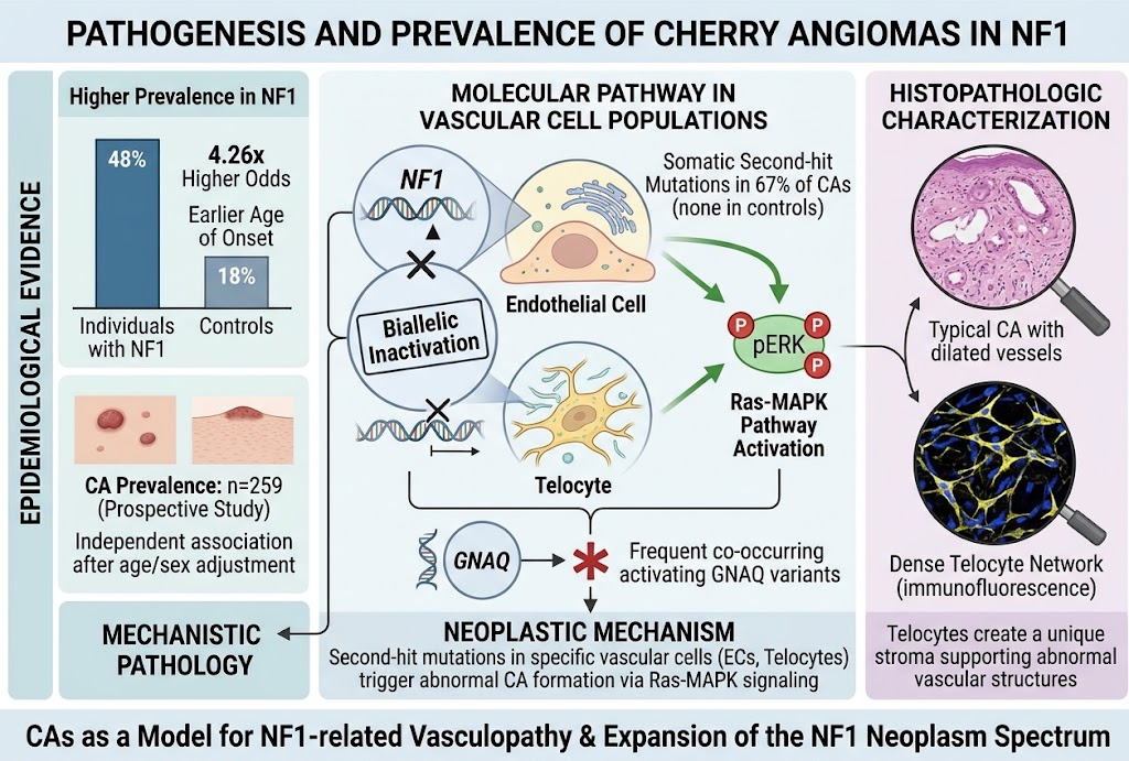
The study found a significantly higher prevalence of cherry angiomas in the NF1 group compared to controls: 48% versus 18%, respectively (OR 4.26; 95% CI, 2.44-7.56). Notably, CAs appeared at younger ages in NF1 patients, supporting a disease-specific vascular manifestation rather than age-related skin changes. Propensity score matching adjusted for confounders confirmed the robustness of this association.
Histopathological analyses of NF1-associated CAs showed typical lobular capillary proliferations without atypia or malignancy, consistent with classic cherry angiomas.
Molecular investigations revealed somatic NF1 loss-of-function second-hit variants in 67% (26/39) of NF1-associated CAs but none in control samples, indicating biallelic NF1 inactivation is a key pathogenic event in NF1-related CAs. Comprehensive genomic profiling also uncovered frequent concurrent activating mutations, predominantly in GNAQ, which has been implicated in vascular tumors and capillary malformations.
Cell-specific sequencing highlighted that NF1 second hits were primarily localized to endothelial cells and telocytes — specialized interstitial cells involved in vascular homeostasis — with higher variant allele frequencies observed in endothelial populations, indicating their central role in lesion formation. Immunofluorescence staining demonstrated marked upregulation of phosphorylated extracellular signal-regulated kinase (phospho-ERK), reflecting activated Ras-MAPK pathway signaling downstream of NF1 loss.
Expert Commentary
This study provides compelling mechanistic and epidemiologic evidence that cherry angiomas are not incidental skin findings but represent a previously unrecognized vascular manifestation of NF1. The biallelic inactivation of NF1 within vascular endothelial cells parallels the ‘‘two-hit’’ hypothesis previously established for NF1-associated tumors, emphasizing the tumor suppressor role of NF1 in vasculature. The frequent co-mutation in GNAQ may further promote endothelial proliferation and angiogenesis.
From a clinical standpoint, recognizing CAs as part of the NF1 spectrum may improve earlier diagnosis and surveillance of vascular complications in NF1 patients. The robust molecular characterization advances understanding of NF1 vasculopathy and opens avenues for targeted therapeutics, particularly inhibitors of the Ras-MAPK axis.
Limitations include the cross-sectional design, restricting inference of lesion evolution over time, and sampling primarily from one tertiary center, which may affect generalizability. Longitudinal cohort studies are warranted to determine causality, lesion progression, and clinical outcomes. Additionally, the influence of other genetic or environmental factors on CAs in NF1 requires exploration.
Conclusion
This study elucidates cherry angiomas as a frequent vascular cutaneous manifestation in Neurofibromatosis Type 1, supported by epidemiological enrichment and molecular evidence of NF1 biallelic inactivation in vascular endothelial cells. These findings extend the spectrum of NF1-associated neoplasms beyond classic neurofibromas to include vascular proliferations, offering new insights into NF1 pathobiology and vasculopathy. Recognition of CAs in NF1 patients should inform clinical evaluation and stimulate research into targeted interventions for NF1-related vascular lesions.
Funding and Clinical Trial Registration
The study was conducted at the Henri-Mondor University Hospital with no specific external funding declared in the provided content. Clinical trial registration details were not specified.
References
Fertitta L, Pasmant E, Bergqvist C, et al. Cherry Angiomas in Individuals With Neurofibromatosis Type 1. JAMA Dermatol. 2026; Published online August 19, 2026. PMID: 42616496.
National Institutes of Health. Neurofibromatosis Type 1 information page. https://www.ninds.nih.gov/health-information/disorders/neurofibromatosis-type-1
Upadhyaya M, Cooper DN. Neurofibromatosis Type 1: Molecular and Cellular Biology. Int J Mol Sci. 2019;20(4):1003.
Bayat A, et al. GNAQ Mutations and the Ras-MAPK Pathway in Vascular Anomalies. Int J Mol Sci. 2020;21(14):5034.
Riccardi VM. Neurofibromatosis: Phenotype, Natural History, and Pathogenesis. 3rd ed. Johns Hopkins University Press; 1992.