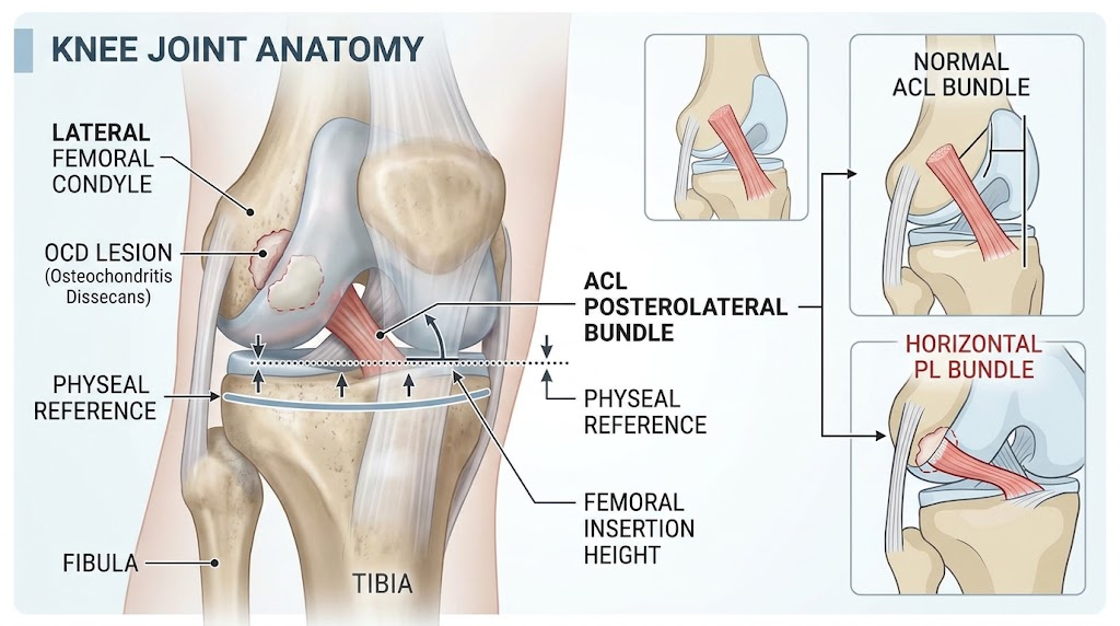
Osteochondritis dissecans (OCD) of the lateral femoral condyle (LFC) associates with a significantly more horizontal position of the ACL posterolateral (PL) bundle insertion compared to controls.
The study used MRI-based morphometric measurements of the PL bundle relative to physis and femoral condyle height in pediatric patients aged 8 to 16 years.
A more distal femoral insertion point of the PL bundle may represent an independent anatomical factor for OCD development and influence lesion healing potential.
Study Background
Osteochondritis dissecans (OCD) is a joint condition characterized by focal subchondral bone necrosis and fragmentation which often involves the femoral condyles of the knee. Pediatric and adolescent populations are predominantly affected, and the disease manifests with pain, swelling, and impaired joint function. Among various hypothetical etiologies, including repetitive microtrauma, ischemia, and genetic predisposition, anatomical factors have remained insufficiently explored. The anterior cruciate ligament (ACL), particularly its posterolateral (PL) bundle, plays a vital biomechanical role in knee stability, and its anatomical relationship with adjacent articular surfaces may influence OCD pathogenesis. Previous investigations have not specifically examined how the ACL’s orientation correlates with OCD lesions in the lateral femoral condyle (LFC). Understanding these anatomical nuances could inform prognosis and therapeutic approaches in clinical pediatric orthopedics.
Study Design
This retrospective case-control study included 90 knees from 90 pediatric patients (mean age 12.5 years, range 8–16 years; 67% male), divided evenly into three cohorts matched by age and sex. Group 1 consisted of patients with OCD localized to the lateral femoral condyle (LFC). Group 2 included patients with OCD of the medial femoral condyle (MFC), serving as a lesion-specific control group. Group 3 comprised individuals with no structural knee pathology as healthy controls.
Inclusion criteria mandated patients below 18 years, with intact ACLs and no prior knee surgeries. Patients with discoid meniscus were excluded to eliminate confounding anatomical variations. Magnetic resonance imaging (MRI) was used to perform detailed morphometric analysis. On coronal MRI slices, the vertical height of the ACL PL bundle insertion was measured relative to the physeal height and the LFC height, serving as normalization parameters. Additionally, the femorotibial angle was calculated to ensure consistent measurement planes among subjects.
Key Findings
The study revealed a statistically significant difference in the vertical height of the PL bundle insertion between the groups. Specifically, the PL bundle insertion in patients with lateral femoral OCD (Group 1) was positioned more distally (more horizontal ACL orientation) compared to both OCD of the medial femoral condyle (Group 2) and controls (Group 3).
Quantitatively, the mean relative PL bundle height compared to femoral condyle height was 0.36 (SD 0.06) in Group 1 versus 0.43 (SD 0.09) in Group 2 and 0.43 (SD 0.08) in controls. When referenced to physeal height, values were 0.46 (SD 0.07) for Group 1, 0.50 (SD 0.11) for Group 2, and 0.55 (SD 0.11) for controls. These results were statistically significant with P < .001.
The data indicate that the posterolateral ACL bundle attaches more distally on the lateral femoral condyle in patients with OCD of this region. This anatomical variation suggests a biomechanical environment distinct from that encountered in medial femoral condyle OCD or healthy knees.
Expert Commentary
This study provides novel insights into the anatomical factors potentially influencing OCD of the lateral femoral condyle. The distal (more horizontal) insertion of the posterolateral bundle may alter knee joint biomechanics, potentially increasing stress on the subchondral bone of the lateral condyle and contributing to lesion formation or impaired healing. Although previous research did not directly connect ACL bundle positioning to OCD, the current findings are biologically plausible given the ACL’s role in stabilizing rotational forces.
However, limitations include the retrospective design and reliance on indirect morphometric measures. The study population, though well-matched, was limited in size, and factors such as activity level, biomechanical loading patterns, or genetic predispositions were not controlled. Future prospective studies should integrate dynamic biomechanical assessments and longitudinal follow-up to clarify causality and prognostic significance of the PL bundle positioning.
Conclusion
The identified association between a more horizontal posterolateral ACL bundle position and OCD of the lateral femoral condyle underscores an anatomical contributor to this pediatric orthopedic pathology. This distal femoral attachment point variation might serve as an independent predictor of OCD lesion healing outcomes and could inform tailored interventions. Future investigative efforts should aim to elucidate mechanistic pathways linking ACL morphology to OCD pathogenesis and test whether interventions addressing such biomechanical factors can enhance therapeutic success.
Funding and Clinical Trials
The original study does not report specific funding sources or registration on clinical trial databases.
References
1. Lehoczky G, Khoo B, Lin Z, Shea KG. More Horizontal ACL Posterolateral Bundle Position in Osteochondritis Dissecans of the Lateral Femoral Condyle. Am J Sports Med. 2026 Sep;54(12):2989-2995. PMID: 42745400.
2. Wall EJ, Hypolite IO. Osteochondritis dissecans of the knee: current concepts and future directions. J Am Acad Orthop Surg. 2007 Apr;15(4):211-21. doi:10.5435/00124635-200704000-00004.
3. Steiner ME, Tasto JP. Anterior cruciate ligament anatomy and surgical reconstruction techniques. Sports Med Arthrosc Rev. 1995;3(2):75-82.
4. McDermott ID, Schiller JR, Amis AA. Biological and biomechanical considerations in osteochondritis dissecans of the knee. J Bone Joint Surg Br. 1993 Sep;75(5):682-6.