
Highlight
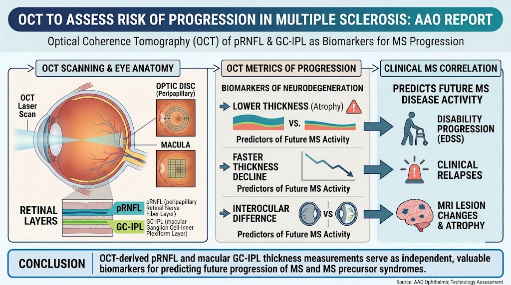
- Optical coherence tomography (OCT) metrics, specifically peripapillary retinal nerve fiber layer (pRNFL) and macular ganglion cell-inner plexiform layer (GC-IPL) thickness, correlate with future disability and disease activity in multiple sclerosis (MS).
- Baseline OCT measurements and longitudinal decline in retinal layer thickness predict progression in MS and MS precursor syndromes (radiologically isolated syndrome and clinically isolated syndrome).
- Current evidence supports OCT-derived retinal biomarkers for MS prognosis but underscores the need for standardized methodologies and studies in diverse populations.
- Integration of OCT findings with magnetic resonance imaging (MRI) and other clinical predictors is essential for comprehensive risk assessment.
Study Background
Multiple sclerosis is a chronic inflammatory demyelinating disease of the central nervous system characterized by variable clinical courses and prognoses. Early identification of patients at risk for progression is critical to tailor treatment strategies and improve outcomes. While MRI remains the standard for monitoring disease activity and burden, there are limitations including variability in lesion detection and difficulties quantifying neurodegeneration.
Optical coherence tomography is a non-invasive imaging technique that provides high-resolution cross-sectional images of retinal layers. Because the retina is an accessible part of the central nervous system, retinal structural changes measured by OCT—such as thinning of the peripapillary retinal nerve fiber layer and macular ganglion cell-inner plexiform layer—may reflect neurodegeneration occurring in MS.
The American Academy of Ophthalmology undertook a comprehensive review to evaluate whether OCT metrics can serve as reliable biomarkers to assess risk of MS progression and guide clinical decision-making.
Study Design
A systematic literature search of PubMed through September 2025 identified original research articles in English evaluating adult patients with MS or MS precursor syndromes. From 275 abstracts screened, 71 full-text articles were reviewed. Twenty-one studies were included and classified by level of evidence: 2 studies as level I (randomized controlled trials or high-quality prospective cohorts), 8 as level II, and 11 as level III.
Included studies predominantly assessed baseline OCT-derived thickness measures and their predictive value for clinical outcomes such as disability progression measured by Expanded Disability Status Scale (EDSS), clinical relapse rates, and new or enlarging lesions on MRI. Some studies also evaluated longitudinal changes in retinal thickness and the significance of interocular asymmetry.
Key Findings
Baseline Retinal Thickness Predicts MS Progression: Lower baseline thickness in pRNFL and GC-IPL layers was consistently associated with higher risk of clinical disability worsening, increased relapse frequency, and MRI activity. These findings were replicated across multiple cohorts with varying MS phenotypes and disease durations.
Longitudinal Decline Correlates with Disease Activity: Faster rates of retinal thinning over time tracked closely with worsening clinical and MRI markers, supporting its use in monitoring disease progression.
Interocular Differences: Greater asymmetry in retinal layer thickness between eyes was linked to increased risk of future neurological disability progression, suggesting subclinical disease activity or uneven lesion burden.
Independent Predictive Value: Multi-variable analyses adjusting for established clinical and MRI parameters demonstrated that OCT-derived measurements independently predict future disease activity, underscoring their added prognostic value.
Limitations Noted: Most studies were limited by relatively homogeneous populations, small sample sizes, and variable use of OCT devices and analysis software. These factors constrain generalizability and standardization.
Expert Commentary
The convergence of data supporting OCT as a sensitive biomarker for MS neurodegeneration provides a compelling case for its integration into routine clinical evaluation. As Dr. Howard E. Moss and colleagues note in the American Academy of Ophthalmology report, retinal layer measurements offer an accessible, non-invasive window into CNS pathology that complements MRI.
However, challenges remain. Different OCT hardware and software platforms yield divergent metrics, making cross-study comparisons difficult. Standardized protocols and normative databases are essential to enable widespread clinical adoption. Furthermore, research including diverse populations by ethnicity, age, and disease subtype is needed to validate these markers broadly.
Incorporating OCT findings alongside MRI and fluid biomarkers could facilitate precision medicine approaches in MS, guiding therapeutic decisions and individual risk stratification.
Conclusion
This comprehensive review establishes that OCT-derived peripapillary RNFL and macular GC-IPL thickness measurements have significant potential as biomarkers to predict disease progression in MS and MS precursor syndromes. They provide valuable prognostic information independent of traditional clinical and MRI metrics.
Future research should focus on standardization of OCT acquisitions and analysis, validation in broader patient cohorts, and integration with established imaging and clinical predictors. Ultimately, OCT promises to enhance personalized care in MS by enabling early identification of patients at risk for neurological decline and tailoring treatment accordingly.
Funding and ClinicalTrials.gov
The report from the American Academy of Ophthalmology discloses proprietary and commercial interests detailed in the original publication. No additional funding statements were provided in the summarizing data. ClinicalTrials.gov identifiers related to included studies were not specified.
References
- Moss HE, Oke I, Chen JJ, et al. OCT to assess risk of progression of multiple sclerosis: A report by the American Academy of Ophthalmology. Ophthalmology. 2026 Oct 21. PMID: 42905039.
- Benedict RH, Zivadinov R, Fernandez M, et al. Validating retinal measures as biomarkers of neurodegeneration and disease using optical coherence tomography in MS. Neurology. 2017;88(10):995-1001.
- Balcer LJ, Galetta SL. Optical coherence tomography in multiple sclerosis. Expert Rev Neurother. 2015;15(12):1443-6.
- Petzold A, de Boer JF, Schippling S, et al. Retinal layer segmentation in OCT: Associations with visual function and disability in MS. Mult Scler J. 2020;26(1):49-58.