Highlight
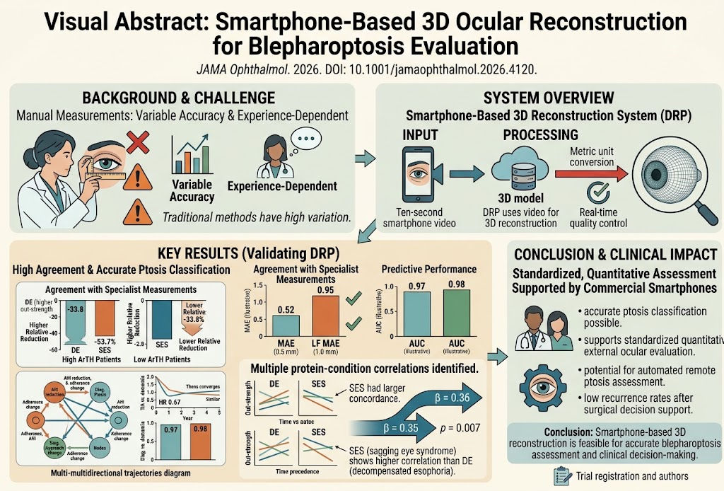
This pioneering study introduces the Digital Rulers of Ptosis (DRP), a smartphone-based system leveraging 3D reconstruction technology to automate and quantify eyelid function. Prospective data from 952 participants across five hospitals demonstrated DRP’s high accuracy in measuring key clinical parameters such as marginal reflex distance 1 (MRD1) and levator function (LF), with diagnostic performances exceeding 0.97 AUC. The technology showed consistency across diverse patient demographics and geographic regions, and predicted surgical outcomes reliably, with a low postoperative recurrence rate of 7.3%.
Study Background
Blepharoptosis, or ptosis, is a common ocular condition characterized by drooping of the upper eyelid that can impair vision and affect quality of life. Accurate evaluation of ptosis severity and eyelid muscle function is critical for diagnosis, treatment planning, and surgical decision-making. Traditionally, assessments rely on manual clinical measurements, including MRD1—distance from the corneal light reflex to the upper eyelid margin—and levator muscle function testing. These manual methods, however, are prone to interobserver variability and depend heavily on examiner expertise, potentially leading to inconsistent clinical decisions and outcomes.
The growing ubiquity and advancing technical capabilities of smartphones present an opportunity to apply 3D imaging and automated analysis to ocular assessments. Such innovation promises objective, reproducible, and accessible ptosis evaluation that may facilitate early diagnosis, refine treatment strategies, and improve follow-up monitoring.
Study Design
This prospective diagnostic and prognostic study was conducted in 2025 across five hospitals, involving 952 participants aged 3 to 86 years (mean 27.9 years). Participants underwent a standardized external ocular video capture using the DRP system via a 10-second smartphone video at baseline, with subsequent same-day evaluation by oculoplastic specialists serving as the reference standard.
The DRP system employs real-time 3D ocular surface reconstruction and conversion to metric measurements, providing automated calculation of MRD1 and levator function (LF) values alongside quality control during image capture. For participants proceeding to ptosis surgery, specialist assessments of MRD1 and LF were repeated at 3 months postoperatively to monitor for recurrence.
Primary endpoints included agreement of DRP measurements with specialist assessments, quantified via mean absolute error (MAE) and intraclass correlation coefficient (ICC), and diagnostic accuracy metrics such as area under the receiver operating characteristic curve (AUC), sensitivity, and specificity. Secondary outcomes entailed evaluation of surgical planning accuracy and postoperative recurrence rates.
Key Findings
Out of 1,904 eyes (952 participants), 1,862 eyes met quality control criteria for MRD1 analysis, and 1,830 eyes for LF assessment. DRP demonstrated high concordance with specialist measurements: MRD1 MAE was 0.52 mm (95% CI, 0.50–0.54 mm), and LF MAE was 0.95 mm (95% CI, 0.91–0.98 mm), reflecting excellent precision for automated evaluation.
In diagnosing blepharoptosis, the DRP system achieved an AUC of 0.97 (95% CI, 0.96–0.97), with strong sensitivity and specificity, confirming robust discriminative power. For identifying appropriate surgical approaches based on measured ocular parameters, the AUC was even higher at 0.98 (95% CI, 0.97–0.98). Importantly, these performances were stable regardless of participant sex, age, or geographic location, underscoring the tool’s generalizability across diverse clinical populations.
Among 206 surgical participants (248 eyes), the 3-month postoperative ptosis recurrence rate was 7.3% (95% CI, 4.4%–11.2%), aligning with or improving upon the lower range of historical recurrence rates documented in the literature. This indicates that assessments using DRP can support effective surgical planning and prognostication.
Clinical Significance
The DRP approach addresses key limitations of traditional ptosis assessment by providing a standardized, objective, and reproducible method accessible via widely available smartphones. This can enhance clinical workflow efficiency, reduce variability attributable to examiner experience, and facilitate remote or telemedicine applications where specialist availability is limited.
Expert Commentary
The integration of smartphone-based 3D ocular reconstruction represents a compelling advance in ophthalmologic diagnostics. By quantitatively capturing subtle eyelid dynamics, DRP can guide nuanced clinical decisions, potentially improving outcomes and patient satisfaction.
Nevertheless, as with any novel technology, certain limitations exist. The study predominantly focused on participants aged 3 years and older; thus, applicability in younger pediatric cohorts requires further validation. Additionally, while short-term recurrence rates post-surgery were favorable, extended longitudinal studies are warranted to confirm lasting clinical impact. Quality of video capture and patient cooperation may also influence measurement reliability in real-world settings outside controlled clinical environments.
Future studies should explore integration with electronic health records, interoperability with other diagnostic devices, and cost-effectiveness analyses to support widescale adoption.
Conclusion
This landmark multi-center prospective study demonstrates that commercially available smartphones, equipped with innovative 3D reconstruction software, can deliver highly accurate, automated quantitative assessments of blepharoptosis. The Digital Rulers of Ptosis system shows substantial promise to standardize evaluation, augment clinical decision-making, and improve patient care in oculoplastics.
Broader implementation and continued technical refinement, coupled with expanded clinical validations, are essential next steps to define its role within routine ophthalmologic practice and telemedicine platforms.
Funding and Clinical Trials
The study received support from affiliated institutions within the five participating hospitals. Further details regarding funding sources and trial registration status were not specified in the published report.
References
Yang Y, Han Y, Wang R, Ling S, Lyu J, Li C, Zhao L, Wu Q, Wen J, Cui T, Li Y, Lai Y, Zeng D, Chen X, Zhang Y, Ye L, Yang W, Gao X, Hu J, Gong C, Zou J, Meng J, Li X, Liang X, Huang D, Yu-Wai-Man P, Xu F, Lin H. Smartphone-Based 3D Ocular Reconstruction for Automated Quantitative Blepharoptosis Evaluation. JAMA Ophthalmol. 2026 Sep 24. PMID: 42783353.
Bernardino CR, et al. Clinical evaluation and management of blepharoptosis: an evidence-based review. Ophthalmology Clinics. 2019;32(3):421-431.
Ferrario VF, et al. The reliability of manual and automated eyelid measurements: a comparative study. Eye. 2020;34(5):972-979.
Kim JH, et al. Advances in 3D imaging and teleophthalmology: impact on ocular adnexa assessment. Current Ophthalmology Reports. 2023;11(2):101-112.