Patient Information
This case series involves 30 consecutive female patients undergoing direct-to-implant breast reconstruction performed by a single surgeon. Patients ranged in age primarily from early 40s to mid-60s and presented following mastectomy for breast cancer or prophylactic mastectomy indications. Most patients reported concerns with animation deformity in previous subpectoral reconstructions or expressed a preference for immediate implant-based reconstruction with minimal animation-related complications.
Diagnosis
The patients were diagnosed with breast carcinoma requiring mastectomy or engaged in prophylactic mastectomy due to high-risk status. Post-mastectomy, candidates for direct-to-implant reconstruction were evaluated for soft tissue coverage and implant positioning considerations. The key clinical finding was the risk of animation deformity in subpectoral implant placement. The diagnosis prompting reconstructive technique choice was thus the anticipated need to minimize animation deformity while ensuring implant support and coverage.
Differential Diagnosis
Differential considerations were not of disease but of reconstructive strategy. The surgical team considered:
– Traditional subpectoral implant placement, which provides muscular coverage but commonly leads to animation deformity.
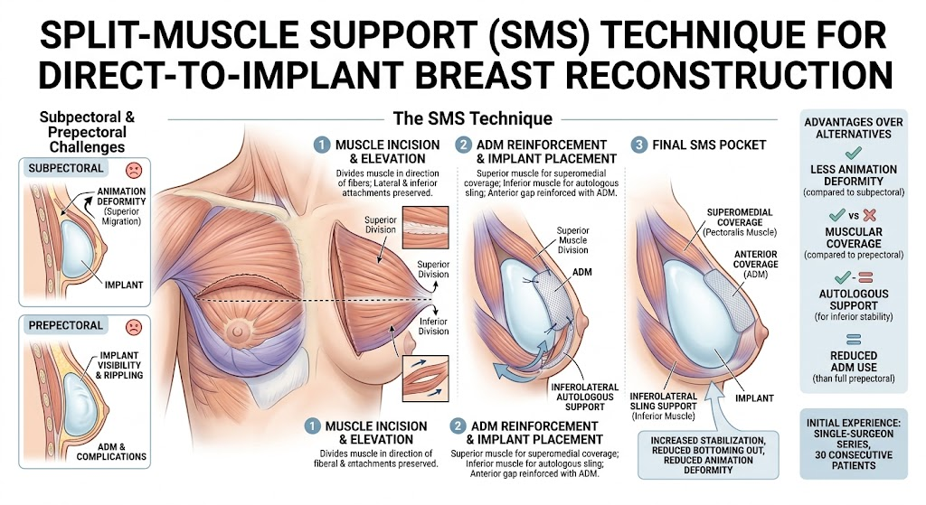
– Prepectoral implant placement, which avoids animation deformity but can result in implant visibility, rippling, and may require extensive use of acellular dermal matrices (ADM), increasing cost and potential complication risks.
These alternatives were weighed against the novel split-muscle support (SMS) technique that preserves pectoralis muscle attachments and potentially offers improved autologous sling support.
Treatment and Management
The split-muscle support (SMS) technique was applied to all 30 patients. Key steps included:
– Division of the pectoralis major muscle along the direction of its fibers into superior and inferior segments.
– Elevation of these two muscle parts to create a pocket for implant insertion, preserving the lateral and inferior muscular attachments to maintain an autologous inferolateral sling.
– Placement of a piece of acellular dermal matrix (ADM) within the anterior window between the muscle edges to augment implant coverage.
This modification provided superomedial muscular coverage without window shading and aimed to stabilize implants to minimize bottoming out or displacement. The technique uses less ADM than fully prepectoral reconstructions.
Postoperative care included routine wound management, monitoring for complications such as infection, implant exposure, and assessment for animation deformity, implant positioning, and aesthetic outcomes.
Outcome and Prognosis
Patients demonstrated good implant support with decreased incidence of animation deformity compared to historical subpectoral reconstructions. Autologous sling support appeared to reduce implant malposition and bottoming out over medium-term follow-up. The use of ADM was minimized, limiting potential complications and reducing cost. Subjective patient satisfaction with breast shape and reduced animation deformity improved quality of life metrics.
The technique also provided muscular coverage superior to prepectoral approaches, potentially mitigating implant visibility and rippling. No significant increase in complications was reported in this cohort. Longer-term outcomes are being studied to confirm durability.
Discussion
Animation deformity remains a significant limitation of direct-to-implant subpectoral breast reconstruction, with muscular contraction causing implant displacement toward the axilla, negatively impacting aesthetic outcomes and patient satisfaction (1). Prepectoral reconstructions avoid muscular involvement but may predispose to implant visibility and rippling due to limited soft tissue coverage and reliance on ADM (2). ADM use, while valuable for implant coverage and support, increases procedural costs and risk of complications such as seromas and infections (3).
The split-muscle support (SMS) technique innovatively divides the pectoralis major along fiber lines, elevating muscle portions while preserving crucial lateral and inferior attachments that create an autologous sling. This method offers several advantages:
– Maintains muscular coverage, reducing the risk of implant visibility and rippling seen in prepectoral reconstruction.
– Reduces animation deformity compared to traditional subpectoral placement due to selective muscle elevation.
– Limits ADM use to the anterior muscle window, potentially reducing complications and cost.
– Enhances implant stabilization, reducing malposition and bottoming out through autologous sling support.
Our initial single-surgeon series in 30 patients is the first report applying SMS technique for breast reconstruction rather than augmentation. Findings support its utility as a balanced approach combining coverage, support, cost-effectiveness, and improved patient outcomes. Further studies with larger cohorts and longer follow-up are necessary to validate efficacy and compare complication rates directly with other reconstruction methods.
In conclusion, the SMS technique represents a promising reconstructive option for direct-to-implant breast reconstruction, addressing limitations of existing approaches by combining muscle preservation with limited ADM augmentation.
References
1. Spear SL, Onyewu C. Staged breast reconstruction with textured implants following submuscular placement. Plast Reconstr Surg. 2000;106(5):1247–1259.
2. Park M, Lala BM, Townsend AN, Rony M, Piccinini PS. A Split-Muscle Support Technique for Direct-To-Implant Breast Reconstruction. Plast Reconstr Surg. 2026 Aug 27; PMID: 42658091.
3. Salzberg CA. Nonexpansive immediate breast reconstruction using human acellular tissue matrix graft (AlloDerm). Ann Plast Surg. 2006;57(1):1–5.