Highlight
- A novel four-tier categorization of petroclival and temporal bone meningiomas correlates with key surgical outcomes.
- The tier system better predicts extent of tumor resection, complication rates, and tumor recurrence than previous classifications.
- Tumor involvement of critical neurovascular structures defines the highest-risk groups with more complex surgeries.
- This framework may improve preoperative planning and patient-surgeon communication regarding prognosis and risks.
Study Background
Petroclival and temporal bone meningiomas are uncommon skull base tumors that often present clinical challenges due to their complex anatomical relationships. These tumors share some clinical features with vestibular schwannomas but differ significantly in management complexities, especially regarding the involvement of adjacent dura and neurovascular structures. Surgical resection remains a mainstay for symptomatic or progressive lesions; however, approaches are technically demanding because of the tumor’s intricate location near the brainstem, cranial nerves, and major vessels such as the internal carotid artery and jugular bulb.
Existing classification systems for petroclival meningiomas primarily focus on anatomical tumor location or size but lack consistent correlation with surgical outcomes. This hampers surgeons’ ability to anticipate operative difficulty and postoperative risks accurately.
Hence, there is an unmet need for a clinically meaningful descriptive framework that can stratify patients not only by tumor location but also by involvement of critical structures, thereby aiding in outcome prediction, surgical planning, and informed consent.
Study Design and Methods
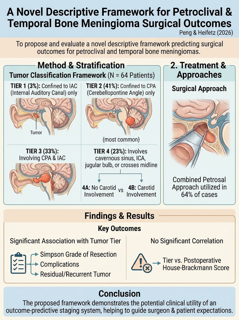
This retrospective analysis reviewed medical records of 64 patients diagnosed with petroclival meningiomas at a tertiary care referral center. Tumors were classified into four tiers based on location and involvement of anatomical structures:
- Tier 1: Tumor confined to internal auditory canal (IAC) only.
- Tier 2: Tumor confined to cerebellopontine angle (CPA) only.
- Tier 3: Tumor involving both CPA and IAC.
- Tier 4: Tumor involving cavernous sinus, internal carotid artery (ICA) or jugular bulb, or crossing midline; subdivided into 4A (no ICA involvement) and 4B (with ICA involvement).
Patients underwent observation, radiation therapy, or surgical resection based on clinical indications. Surgical approaches and outcomes including extent of resection according to Simpson grading, postoperative complications, tumor recurrence or residual disease, and postoperative facial nerve function (House-Brackmann score) were analyzed in relation to tumor tier.
Key Findings
The distribution of patients across tiers was as follows: Tier 2 (41%) was the most frequent, followed by Tier 3 (33%), Tier 4 (23%), and Tier 1 (3%). Among those undergoing surgery, the combined petrosal approach was utilized in 64% of cases, reflecting the anatomical complexity.
Statistically significant associations were found between tumor tier and several critical surgical outcome parameters:
- Simpson Grade of Resection: More extensive tumors (higher tiers) had lower rates of complete resection (Simpson I/II), indicating higher surgical challenge.
- Complication Rates: Higher-tier tumors correlated with increased complication rates, likely reflecting involvement of critical neurovascular structures.
- Residual or Recurrent Tumor: Residual tumor and recurrence rates were significantly higher in Tier 4 tumors, underscoring their prognostic importance.
Interestingly, no significant correlation was noted between tumor tier and postoperative facial nerve function as measured by House-Brackmann scores, suggesting that cranial nerve outcomes may be influenced by other factors beyond tumor extent alone.
Expert Commentary
This study presents a clinically actionable and intuitively clear framework for classifying petroclival and temporal bone meningiomas beyond mere anatomical location. By incorporating major vascular and sinus involvement into tier 4 classifications, the system provides granularity relevant to surgical risk and expected outcomes.
The significant correlation of tiers with resection extent and postoperative complications validates the hypothesis that this framework can serve as a practical predictive tool. It can help neurosurgeons and otolaryngologists in preoperative counseling by setting realistic expectations and tailoring surgical strategies.
Limitations include its retrospective design and relatively small sample size; however, the homogeneous cohort from a tertiary referral center adds value. Prospective validation and comparisons with existing systems will be important next steps.
The lack of correlation between tumor tier and facial nerve outcome invites further investigation into patient-specific factors and microsurgical techniques influencing cranial nerve preservation.
Conclusion
The novel descriptive framework for petroclival and temporal bone meningiomas offers significant promise as an outcome-predictive staging system. Its utilization may optimize surgical planning, guide surgeon-patient discussions on prognosis, and ultimately improve personalized management. Future prospective studies should confirm its generalizability and explore integration into multidisciplinary treatment algorithms.
References
1. Peng KA, Heifetz B. A Novel Descriptive Framework for Petroclival and Temporal Bone Meningioma Surgical Outcomes. The Laryngoscope. 2026 Jun 24;136 Suppl 7:S7-S19. PMID: 42339571.
2. Jefferson GL, Sekhar LN, Samii M. Petroclival meningiomas: surgical anatomy and approaches. Neurosurg Clin N Am. 2020;31(4):557-571.
3. Al-Mefty O. Petroclival meningiomas—the importance of surgical approach and the role of radiosurgery. Neurosurg Focus. 1997;3(6):e5.
4. Simpson D. The recurrence of intracranial meningiomas after surgical treatment. J Neurol Neurosurg Psychiatry. 1957;20(1):22–39.
5. House JW, Brackmann DE. Facial nerve grading system. Otolaryngol Head Neck Surg. 1985;93(2):146-147.