
Highlights
- Condyloma lata presents with consistent, reproducible histopathologic features that distinguish it from other secondary syphilis manifestations and mimickers, such as squamous cell carcinoma.
- Recognition of characteristic clinical and histologic patterns—including verrucous plaques, pseudoepitheliomatous hyperplasia, lichenoid lymphoplasmacytic infiltrate, and abundant spirochetes—is critical to accurate diagnosis.
- Extragenital condyloma lata lesions can be clinically unsuspected and are at risk for misdiagnosis, highlighting the importance of histopathologic evaluation.
- Comprehensive understanding of condyloma lata aids clinicians and pathologists in diagnosing secondary syphilis reliably, even in the absence of systemic cutaneous signs.
Background
Syphilis remains a global public health challenge, characterized by its multisystemic involvement and varied clinical presentations. Secondary syphilis, arising weeks to months after the primary lesion, manifests with polymorphic mucocutaneous lesions due to widespread dissemination of Treponema pallidum. Among these, condyloma lata are moist, wart-like anogenital or occasionally extragenital lesions representing active bacterial proliferation and marked immunologic reaction. Despite their diagnostic significance, condyloma lata are underrecognized histopathologically and can mimic other dermatoses including benign verrucous lesions and malignant entities such as squamous cell carcinoma (SCC). Misdiagnosis may delay appropriate therapy and public health interventions.Key Content
1. Clinical and Histopathologic Characteristics of Condyloma Lata
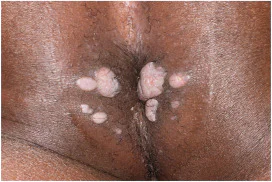
Hyde and Lee (2026) retrospectively reviewed 28 histopathologically confirmed secondary syphilis cases over 15 years; among these, 12 patients with 15 specimens had condyloma lata. The cohort had balanced sex distribution, mean age 37 years, and lesions predominantly localized to anogenital sites but included extragenital locations. Clinically, condyloma lata presented as dome-shaped eroded or verrucous plaques with a distinctive soft, moist appearance, often mistaken for genital warts or SCC, as evidenced by the misdiagnosis in an oral lesion case. Syphilis was clinically suspected in only half of these cases, underscoring diagnostic challenges. Histopathologically, condyloma lata demonstrated a reproducible constellation of features:- Pseudoepitheliomatous hyperplasia: bulbous, irregular, and jagged epidermal hyperplasia mimicking SCC but lacking cytologic atypia typical of malignancy.
- Lichenoid lymphoplasmacytic infiltrate: dense inflammatory infiltrate beneath the epidermis composed predominantly of lymphocytes and plasma cells, consistent with syphilitic immunopathology.
- Prominent neutrophilic exocytosis: neutrophils migrating into epidermis associated with microvesicles showing spongiosis, a sign of active inflammation and epidermal disruption.
- Spirochetes localization: Numerous Treponema pallidum organisms visualized especially in the lower third of the epidermis on Warthin-Starry or immunohistochemical stains, confirming active infection.
2. Histopathologic Variability in Non-Condyloma Lata Secondary Syphilis
The remainder of secondary syphilis cases without condyloma lata morphology showed broad histologic heterogeneity, including psoriasiform, lichenoid, or granulomatous patterns. This variability complicates definitive diagnosis solely on morphology without clinical correlation or spirochetal detection, reinforcing the diagnostic value of recognizing condyloma lata-specific features.3. Diagnostic Challenges and Clinical Implications
The overlap between condyloma lata and entities such as SCC, molluscum contagiosum, and common anogenital warts presents diagnostic pitfalls, especially in extragenital presentations. The frequent absence of additional secondary syphilis signs complicates clinical suspicion. The misdiagnosis of a condyloma lata oral lesion as SCC highlights the potential for overtreatment or delayed syphilis treatment. Integration of detailed histopathologic evaluation with appropriate syphilitic serologies and clinical examination is essential. Awareness of the hallmark histologic features enables timely and accurate diagnosis, facilitating prompt antibiotic therapy and reduction of transmission risk.4. Mechanistic Insights
The localized epidermal pseudoepitheliomatous hyperplasia with irregular acanthosis may represent a reactive pattern to the spirochetes’ presence, while the lichenoid infiltrate reflects a cellular immune response involving plasma cells unique to syphilis. Neutrophilic exocytosis and vesiculation represent acute epidermal injury due to bacterial factors and host inflammation. The concentration of spirochetes in the lower epidermis supports active epidermal invasion and proliferation during condyloma lata formation.Expert Commentary
The study by Hyde and Lee (2026) adds valuable clarity to the histopathologic criteria that reliably distinguish condyloma lata in secondary syphilis. Their rigorous morphologic characterization resolves prior ambiguities hampering diagnosis, especially at less common extragenital sites. The retrospective nature and limited case numbers remain inherent limitations; nonetheless, the reproducibility of key features across 15 specimens lends credibility. Current guidelines recommend a high index of suspicion for syphilis in verrucous mucocutaneous lesions with atypical histology. Immunohistochemical detection of T. pallidum should be pursued when condyloma lata is clinically or morphologically suspected. This work advises against overreliance on clinical impression alone, especially in populations with rising syphilis incidence, including among individuals with HIV co-infection. Future research should expand prospective characterization incorporating molecular diagnostics and explore immunopathologic pathways responsible for pseudoepitheliomatous hyperplasia. Educational initiatives to enhance dermatopathologists’ familiarity with these features could reduce diagnostic delay and prevent unnecessary surgical interventions.Conclusion
Condyloma lata of secondary syphilis represents a distinct clinicopathologic entity with consistent, reliable histopathologic features, including pseudoepitheliomatous hyperplasia, dense lymphoplasmacytic lichenoid infiltrate, neutrophilic exocytosis, and abundant epidermal spirochetes. Recognizing these features is essential for accurate diagnosis, avoiding misdiagnosis as malignant or other benign mimickers, and ensuring appropriate public health management. Enhancing clinical awareness and integrating comprehensive histopathologic and serologic evaluation will improve patient outcomes in syphilis care.References
- Hyde JT, Lee JB. When secondary syphilis is reliably recognizable. J Am Acad Dermatol. 2026 Jun 4;95(4):1067-1070. PMID: 42248252.
- Peeling RW, Mabey D, Kamb ML, Chen XS, Radolf JD, Benzaken AS. Syphilis. Nat Rev Dis Primers. 2017 Jun 8;3:17073. doi:10.1038/nrdp.2017.73. PMID: 28698595.
- Hook EW, Marra CM. Acquired syphilis in adults. N Engl J Med. 1992 Oct 1;327(4):270-8. doi:10.1056/NEJM199207233270406. PMID: 1311766.
- Clark EG, Danbolt N. The Oslo study of the natural history of untreated syphilis. I. The distribution of the stages and the long-term clinical behavior of untreated syphilis. Acta Derm Venereol Suppl (Stockh). 1955;35(34):1-368. PMID: 13221870.
- Singh AE, Romanowski B. Syphilis: Review with emphasis on clinical, epidemiologic, and some biologic features. Clin Microbiol Rev. 1999 Apr;12(2):187-209. doi:10.1128/CMR.12.2.187. PMID: 10223978.
Specialty calculators
For clinical reference; use professional judgment.