Highlight
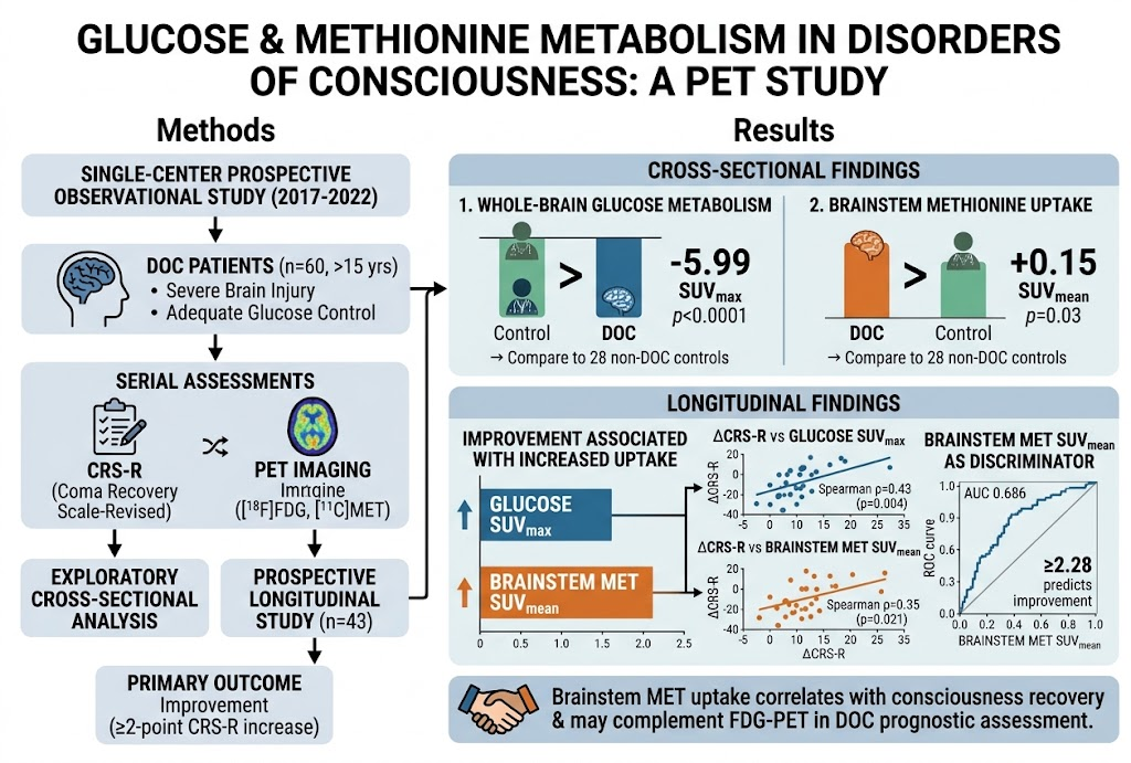
This investigation identifies key metabolic changes in patients with disorders of consciousness (DoC) following severe brain injury by employing both [18F]fluorodeoxyglucose (FDG) and [11C]methionine (MET) PET imaging. Notably, decreased global glucose metabolism contrasts with increased brainstem methionine uptake, the latter correlating with clinical improvement in consciousness. This novel metabolic biomarker could complement FDG-PET in predicting recovery trajectories.
Background: Clinical Context and Disease Burden
Disorders of consciousness (DoC), including vegetative state and minimally conscious state, commonly result from severe brain injury and represent significant clinical challenges. Accurate prognostication is critical yet difficult, as clinical assessments alone have limitations. FDG-PET imaging of glucose metabolism has emerged as a valuable tool for detecting residual brain activity, serving as a marker for conscious processing potential. However, the role of brain amino acid metabolism, especially methionine uptake, has not been thoroughly characterized in this patient population. Understanding additional metabolic pathways could improve prognostic accuracy and guide rehabilitation strategies.
Study Design and Methodology
This single-center, prospective observational study enrolled patients over 15 years old with DoC due to severe brain injury admitted to a Rehabilitation Center from 2017 to 2022. Patients had stable glucose control to minimize metabolic confounders. The study comprised both a cross-sectional evaluation at baseline (average 20.9 months post-injury) and a longitudinal component tracking clinical and metabolic changes over time. Serial assessments using the Coma Recovery Scale-Revised (CRS-R) quantified consciousness levels, with ≥2-point CRS-R increase defining improvement. PET imaging included FDG for glucose metabolism and MET for methionine uptake. Primary analyses evaluated correlations between metabolic PET metrics (SUVmax for glucose, SUVmean for methionine) and CRS-R changes using Spearman correlation, supplemented by logistic regression and receiver operating characteristic curve analyses to explore discriminatory power.
Key Findings
Sixty patients formed the cross-sectional cohort compared against 28 non-DoC controls, and 43 patients entered the longitudinal analysis. Compared with controls, DoC patients exhibited significantly reduced whole-brain glucose metabolism (SUVmax mean difference -5.99, p < 0.0001), reflecting widespread cortical hypometabolism. Conversely, brainstem methionine uptake was significantly higher (SUVmean mean difference 0.15, p = 0.03), indicating a possible compensatory or reparative metabolic response.
During follow-up, 49% demonstrated clinical improvement with a median CRS-R increase of 4 points. Improved patients were on average 49.6 years old, with 33% female, and had baseline median CRS-R of 11. Longitudinal analyses revealed that increased glucose SUVmax and elevated brainstem MET SUVmean correlated positively with CRS-R improvements (Spearman’s ρ = 0.43, p = 0.004 for glucose; ρ = 0.35, p = 0.021 for methionine). Notably, a brainstem MET SUVmean ≥2.28 had moderate accuracy in discriminating patients who improved (AUC 0.686), supporting its prognostic potential.
Expert Commentary
This study introduces a novel dimension to metabolic imaging in DoC by integrating methionine PET with standard FDG-PET. The finding of increased brainstem methionine uptake associated with clinical improvement suggests enhanced amino acid metabolism or protein synthesis pathways in brainstem regions may underpin recovery mechanisms. These insights complement existing knowledge emphasizing cortical glucose metabolism deficit as a hallmark of DoC. Although promising, results remain exploratory given the moderate sample size, single-center design, and inherent heterogeneity in brain injuries and rehabilitation courses.
Clinical application will require larger, multi-institutional validations and mechanistic studies elucidating the biological basis of altered methionine metabolism. Moreover, integrating PET metabolic markers with electrophysiologic and neurobehavioral assessments could enhance multidimensional prognostication. This metabolic profiling could also open avenues for targeted metabolic modulation in therapeutic interventions.
Conclusions and Future Directions
The study compellingly demonstrates that combined PET imaging of glucose and methionine metabolism provides complementary information in patients with disorders of consciousness after brain injury. Decreased global glucose metabolism remains a robust marker of impaired brain function, whereas elevated brainstem methionine uptake emerges as a novel correlate of consciousness recovery. Methionine PET may thus augment current metabolic assessment protocols and assist clinicians in prognostication and monitoring.
Future research should aim at validating these findings in larger cohorts, dissecting underlying molecular mechanisms, and exploring implications for personalized rehabilitation and therapeutic targeting. Enhanced understanding of cerebral amino acid metabolism post-injury may pave the way for novel biomarkers and interventions to improve outcomes in this challenging patient population.
Funding and Registration
The study was registered with the Japan Registry of Clinical Trials (identifier: jRCTs031180091) on January 21, 2019. Funding sources were not detailed in the abstract.
References
- Yamaki T, Oka N, Higuchi Y, Ito D, Kobayashi S. Glucose and Methionine Metabolism in Disorders of Consciousness After Brain Injury: An Exploratory Longitudinal PET Study. Neurology. 2026;107(5):e218419. PMID: 42555891.
- Laureys S, Celesia GG, Cohadon F, et al. Unresponsive wakefulness syndrome: a new name for the vegetative state or apallic syndrome. BMC Med. 2010;8:68. doi:10.1186/1741-7015-8-68.
- Stender J, Mortensen KN, Thibaut A, et al. The minimal energetic requirement of sustained consciousness after brain injury. Brain. 2016;139(Pt 7):2081-2089. doi:10.1093/brain/aww131.
- Stender J, Gosseries O, Bruno M-A, et al. Diagnostic precision of PET imaging and functional MRI in disorders of consciousness: a clinical review. Lancet Neurol. 2014;13(11):1094-1105. doi:10.1016/S1474-4422(14)70170-1.