Introduction
Persistent low-level human chorionic gonadotropin (hCG) following gestational trophoblastic disease (GTD) presents a clinical conundrum regarding prognosis and management. Differentiating benign persistence of low-level real hCG from active gestational trophoblastic neoplasia (GTN) is critical to avoid overtreatment and associated morbidities. The study by Alevato et al. provides a comprehensive retrospective analysis delineating the natural history of persistent low-level real hCG and identifying clinical and biochemical predictors of subsequent GTN, with implications for surveillance strategies in a high-volume referral setting.
Clinical Context and Disease Burden
GTD encompasses a spectrum of disorders arising from placental trophoblastic tissue, ranging from benign hydatidiform moles to malignant GTN. Serial monitoring of serum hCG is pivotal in post-evacuation surveillance due to its high sensitivity as a tumor marker. However, low-level hCG persistence (defined in this study as ≤100 IU/L for ≥3 months) after remission creates diagnostic and therapeutic challenges. Persistent low-level hCG may reflect residual benign trophoblastic elements or early malignancy, necessitating nuanced clinical judgment to optimize patient outcomes while reducing unnecessary interventions.
Study Design and Methods
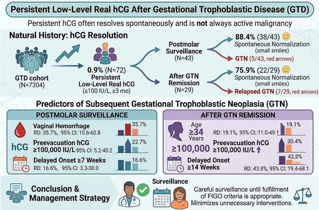
This retrospective cohort study involved 7,304 GTD patients treated at specialized GTD referral centers in Rio de Janeiro, Brazil, between 2000 and 2022. Among these, 72 patients (0.9%) exhibited persistent low-level real hCG, either during postmolar surveillance or after achievement of GTN remission. Persistent low-level hCG was defined as sustained serum hCG ≤100 IU/L over a minimum of 3 months. Clinical parameters, hCG trajectories, and oncologic outcomes were collected. The analysis utilized risk differences (RD) with 95% confidence intervals (CI) to evaluate associations between clinical features and progression to subsequent GTN.
Key Findings
The majority of patients with persistent low-level real hCG experienced spontaneous normalization: 88.4% (38/43) in the postmolar surveillance subgroup and 75.9% (22/29) after GTN remission. These findings emphasize that persistent low-level hCG does not uniformly signify active malignancy.
However, risk factors for developing GTN differed between patient subgroups:
– Postmolar surveillance group:
– Vaginal hemorrhage increased GTN risk by 35.7% (95% CI: 10.6–60.8).
– Preevacuation hCG ≥100,000 IU/L was associated with a 22.7% increased risk (95% CI: 5.2–40.2).
– Delayed onset of persistent low-level real hCG ≥7 weeks conferred a 16.6% higher risk (95% CI: 3.3–30.0).
– Post-GTN remission group:
– Age ≥34 years elevated relapse risk by 19.1% (95% CI: 11.0–49.1).
– Preevacuation hCG ≥100,000 IU/L increased risk by 30.4% (95% CI: 11.6–49.2).
– Delayed onset of persistent low-level hCG ≥14 weeks conferred the highest increased risk at 43.8% (95% CI: 19.4–68.1).
These findings underscore the importance of individualized risk stratification based on dynamic and baseline clinical variables.
Clinical Implications and Management
Persistent low-level real hCG after GTD frequently reflects a benign course with spontaneous resolution rather than ongoing malignancy. This supports a strategy of vigilant surveillance in line with FIGO guidelines before initiating aggressive treatment. The identified predictors can refine surveillance intensity and duration, potentially alerting clinicians to earlier intervention in high-risk patients. Avoiding premature chemotherapy or surgery in patients destined for spontaneous remission markedly reduces treatment-related morbidity.
Expert Commentary
The study’s strength lies in its large cohort from a high-volume center with long-term follow-up, yielding robust clinical insights. However, being retrospective, it is subject to inherent biases, including inconsistent measurement intervals and potential confounding factors. The heterogeneity in hCG assay sensitivity and thresholds across institutions must also be considered when extrapolating these findings to diverse clinical settings.
Biologically, persistent low-level hCG may stem from residual trophoblastic cells producing variant or less bioactive isoforms that the assay detects but do not indicate proliferative disease. The delayed onset of persistent hCG as a risk factor suggests that early persistent detection may be less ominous than late emergence, perhaps reflecting initial slow involution versus recurrence.
Incorporating these predictors into risk models alongside clinical and imaging data may improve precision medicine approaches in GTD follow-up.
Limitations and Research Gaps
The retrospective design and single geographical region may limit generalizability. Future prospective studies with standardized hCG assays and uniform follow-up protocols across centers are warranted. Integration of molecular markers and imaging modalities could further delineate malignancy risk.
Summary
Persistent low-level real hCG after gestational trophoblastic disease most often normalizes without intervention, highlighting the importance of conservative surveillance. Risk stratification using clinical features such as vaginal bleeding, preevacuation hCG levels, patient age, and timing of hCG persistence facilitates early identification of patients at increased risk of GTN. Adherence to FIGO criteria coupled with individualized surveillance minimizes unnecessary treatment, improves patient quality of life, and optimizes healthcare resources.
Funding and Trial Registration
No specific funding or clinical trial registration details were reported.
References
1. Alevato R, Braga A, Paiva G, et al. Persistent low-level real human chorionic gonadotropin after gestational trophoblastic disease: Natural history and predictors of subsequent gestational trophoblastic neoplasia. Gynecol Oncol. 2026 Aug 11;212:116-124. PMID: 42579943.
2. FIGO Oncology Committee. FIGO staging for gestational trophoblastic neoplasia 2000. Int J Gynaecol Obstet. 2002 Jan;77(1):285–287.
3. Seckl MJ, Sebire NJ, Berkowitz RS. Gestational trophoblastic disease: current management of hydatidiform mole. BMJ. 2010 Jan 28;341:c5124.
4. Hui P, Cheung A. Diagnostic challenges in gestational trophoblastic disease. Obstet Gynecol Clin North Am. 2016 Mar;43(1):163-176.