Highlight
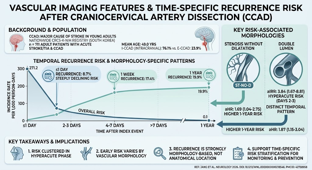
Stroke recurrence after craniocervical artery dissection (CCAD) is heavily concentrated in the hyperacute phase, particularly within the first week after the initial event. Vascular morphology, especially the presence of a double lumen sign and stenosis without dilatation, strongly predicts recurrence risk beyond anatomical dissection location. These findings inform targeted monitoring and secondary prevention strategies in CCAD patients based on imaging morphology and time-specific risk.
Study Background
Craniocervical artery dissection (CCAD) is a significant etiology of ischemic stroke in young adults, representing an important yet understudied contributor to early recurrent stroke risk. Despite its clinical relevance, the temporal pattern of stroke recurrence and the influence of dissection location (intracranial versus extracranial) and vascular morphology on recurrence risk have been poorly delineated. Previous studies suffered from delayed patient enrollment post-index event, limiting insight into the hyperacute risk window. Understanding these dynamics is critical to optimize timely secondary prevention and reduce stroke morbidity.
Study Design
This multicenter cohort investigation utilized the CRCS-K-NIH registry, encompassing a nationwide South Korean stroke population from 2011 to 2021. Adult patients presenting with acute ischemic stroke or transient ischemic attack (TIA) attributed to CCAD diagnosed within 7 days of symptom onset were included (median onset-to-arrival approximately 7.7 hours). Diagnosis was based on vascular imaging demonstrating hallmarks of CCAD. The study principally aimed to characterize 1-year recurrent ischemic stroke risk confirmed by MRI evidencing new or progressive infarction. Recurrence timing was categorized into intervals: ≤1 day, 2-3 days, 4-7 days, and beyond 7 days after the index event. Dissections were anatomically classified as intracranial (I-CCAD) or extracranial (E-CCAD), with detailed imaging morphologies also assessed, including angiographic subtypes and key features such as double lumen presence and stenosis without dilatation.
Key Findings
Among 75,903 acute ischemic stroke or TIA admissions, 711 (0.94%) met criteria for CCAD. The cohort averaged 49 years of age, with women representing 26.9%. Intracranial CCAD accounted for the majority (76.1%). Recurrence kinetics revealed 8.7% stroke recurrence on day 1, escalating to 17.4% within the first week and approaching 20% at 1 year. Incidence rates of recurrence exhibited a sharp decline, from 271.2 events per 1,000 person-days on day 1 to 0.1 events per 1,000 person-days beyond 7 days, underscoring the hyperacute phase as the critical period for secondary events.
Contrary to some prior assumptions, anatomical location of dissection (intracranial versus extracranial) was not independently associated with recurrence risk. However, specific vascular morphologies demonstrated prognostic significance. Stenosis without dilatation conferred a higher recurrence hazard (adjusted hazard ratio [aHR] 1.69, 95% CI 1.04-2.75), and the presence of a double lumen sign was similarly associated with increased 1-year recurrence risk (aHR 1.87, 95% CI 1.15-3.04). Notably, the double lumen sign correlated with a pronounced early recurrence risk between days 2 and 3 post-dissection (adjusted incidence rate ratio 3.84, 95% CI 1.67-8.81).
These observations suggest that vascular morphology contains dynamic prognostic information beyond static anatomical classification, and that recurrence risk is temporally distinct, highest in the first days following onset and diminishing thereafter.
Expert Commentary
The study’s robust, prospective data from a large nationwide registry provide compelling evidence redefining the landscape of stroke recurrence risk after CCAD. The emphasis on morphology, rather than location, as a key determinant aligns with pathophysiological understanding: morphological features such as double lumen likely reflect ongoing intramural hematoma evolution and vessel wall instability, increasing embolic or hemodynamic risk.
These findings facilitate risk stratification enabling clinicians to intensify monitoring and consider aggressive secondary prevention, especially in patients with identified high-risk morphologies during the critical early days. Moreover, the distinction between intracranial and extracranial involvement bears less influence on recurrence likelihood than previously thought, prompting a shift in clinical focus towards vascular imaging phenotypes.
However, some limitations temper extrapolation. The retrospective registry design could introduce selection bias, and the exclusively East Asian population limits generalizability, given possible ethnic or environmental variability in CCAD presentations. Future studies in diverse populations with prospective enrollment immediately after symptom onset would strengthen these conclusions and explore therapeutic implications further.
Conclusion
This comprehensive analysis delineates the temporal clustering of stroke recurrence after CCAD in the early hyperacute phase, with vascular morphology — particularly the double lumen sign and stenosis without dilatation — serving as independent predictors. These insights advocate for morphology- and time-sensitive risk models to guide secondary prevention, potentially reducing recurrent stroke burden in affected young adults. Addressing study limitations with future research in diverse cohorts and interventional trials targeting high-risk morphologies represents a priority opportunity in cerebrovascular medicine.
Funding and Trial Registration
The study was conducted via the CRCS-K-NIH registry in South Korea, covering the period from 2011 to 2021. Detailed funding sources were not specified in the publication. No clinical trial registration number was reported as this is an observational cohort study.
References
Jang SH, Hong JH, Sohn SI, et al. Vascular Imaging Features and Time-Specific Recurrence Risk After Craniocervical Artery Dissection. Neurology. 2026 Sep 18;107(7):e218546. PMID: 42758958.