Highlight
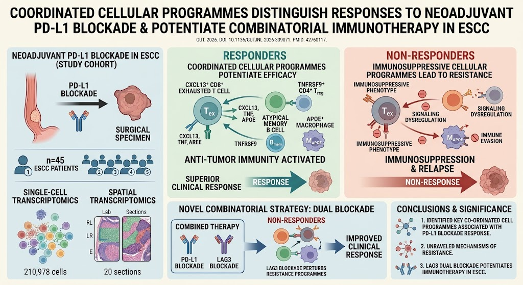
- Identification of coordinated cellular programs driven by CXCL13+ CD8+ exhausted T cells, TNFRSF9+ CD4+ regulatory T cells, atypical memory B cells, and APOE+ macrophages in ESCC response to neoadjuvant PD-L1 blockade.
- Post-treatment non-responders exhibit immunosuppressive cellular programs correlated with therapeutic resistance.
- Lymphocyte activation gene 3 (LAG3) emerges as a promising immunotherapeutic target to potentiate PD-L1 blockade efficacy.
- Comprehensive single-cell and spatial transcriptomic profiling enhances understanding of the tumor immune microenvironment in ESCC.
Study Background
Esophageal squamous cell carcinoma (ESCC) remains a formidable clinical challenge worldwide, characterized by a high mortality rate due to late diagnosis and limited efficacy of conventional treatments. Immune checkpoint blockade, particularly targeting programmed death ligand 1 (PD-L1), has introduced new therapeutic possibilities. Despite advances, response rates to PD-L1 inhibitors in the neoadjuvant setting are variable, and the underlying immunological mechanisms driving clinical outcomes remain incompletely understood. Identifying biomarkers and understanding the cellular interactions governing response or resistance to neoadjuvant PD-L1 blockade could enable precision immunotherapy and improve ESCC patient prognosis.
Study Design
This investigation employed an integrative single-cell transcriptomic analysis of 210,978 cells combined with spatial transcriptomic profiling across 20 ESCC tissue sections to elucidate cellular dynamics underpinning response to neoadjuvant PD-L1 blockade. Patient samples were collected from a clinical trial registered under ChiCTR2400083452. The study objectively compared cellular phenotypes and gene expression profiles at baseline and post-treatment between responders and non-responders, aiming to decode coordinated cellular programs influencing therapeutic efficacy. LAG3 was evaluated as a novel immunotherapeutic target within this context.
Key Findings
The study delineated distinct cellular programs associated with neoadjuvant PD-L1 blockade outcomes. At baseline, responders exhibited coordinated cellular networks comprising CXCL13+ CD8+ exhausted T cells renowned for their antigen-experienced state; TNFRSF9+ (4-1BB+) CD4+ regulatory T cells involved in nuanced immune regulation; atypical memory B cells implicated in adaptive immunity; and APOE+ macrophages correlating with immune modulation. These interacting populations appeared to prime an immune milieu favorable to PD-L1 blockade.
Conversely, post-treatment non-responders manifested a skewed immunosuppressive phenotype within these cellular programs, suggesting a failure to activate effective anti-tumor immune responses. Notably, single-cell and spatial transcriptomic data indicated a reinforced immune evasion landscape preventing therapeutic efficacy in this subgroup.
Importantly, the investigation spotlighted lymphocyte activation gene 3 (LAG3) as an immune checkpoint molecule co-expressed within these coordinated programs. Experimental perturbation of LAG3 pathways demonstrated potential to disrupt immunosuppressive circuits, restoring lymphocyte activity and enhancing PD-L1 blockade response. This finding supports combinatorial immunotherapy approaches integrating LAG3 inhibition with PD-L1 blockade in ESCC.
Expert Commentary
These findings offer seminal mechanistic insights into the immune microenvironmental complexity governing neoadjuvant PD-L1 blockade outcomes in ESCC. The identification of precise immune cell subsets collaborating within a spatially organized tumor microenvironment advances personalized immunotherapy beyond biomarker expression alone. The study’s high-dimensional single-cell and spatial transcriptomics approach represents a cutting-edge methodological benchmark for dissecting tumor-immune interplays.
Nonetheless, while the data strongly indicate LAG3 as a promising combinatorial target, clinical validation in randomized trials is essential. Additionally, understanding the influence of tumor heterogeneity and the broader systemic immune context will be critical for translating these insights into durable therapeutic gains. The study underscores the relevance of integrated multi-omics profiling to expand treatment options for ESCC, a historically intractable malignancy.
Conclusion
This comprehensive analysis reveals that coordinated immune cell programs involving exhausted T cells, regulatory T cells, B cells, and macrophages fundamentally determine ESCC patient response to neoadjuvant PD-L1 blockade. The immunosuppressive phenotype characterizing non-responders highlights mechanistic resistance pathways. Targeting LAG3 within these cellular programs potentiates immunotherapy efficacy, paving the way for innovative combination strategies. These insights provide a framework for individualized immunotherapy development aiming to overcome resistance and improve survival outcomes in ESCC.
Funding and Trial Registration
The study was registered with the Chinese Clinical Trial Registry (ChiCTR2400083452). Specific funding details were not disclosed in the abstract but are typically acknowledged in the full publication.
References
1. Li S, Guo J, Ma J, et al. Coordinated cellular programmes distinguish responses to neoadjuvant PD-L1 blockade and potentiate combinatorial immunotherapy in ESCC. Gut. 2026 Sep 18; PMID: 42760117.
2. Smyth EC, Lagergren J, Fitzgerald RC, et al. Oesophageal cancer. Nat Rev Dis Primers. 2017 Nov 16;3:17048.
3. Chen L, Han X. Anti-PD-1/PD-L1 therapy of human cancer: past, present, and future. J Clin Invest. 2015 Sep;125(9):3384-91.
4. Pauken KE, Wherry EJ. Overcoming T cell exhaustion in infection and cancer. Trends Immunol. 2015 Apr;36(4):265-76.