
Highlight
Histiocytic neoplasms such as Erdheim-Chester disease (ECD), Langerhans cell histiocytosis (LCH), and Rosai-Dorfman-Destombes disease (RDD) frequently involve the nervous system, particularly spinal cord and nerve roots. This retrospective cohort study elucidates characteristic clinical and MRI patterns of such involvement across these diseases. Parenchymal spinal cord infiltration predominates in ECD, while nerve root engagement is more common in RDD. Neurologic disability at presentation is substantial, but treatment yields clinical and radiographic improvement in the majority, especially in RDD and LCH. Early spinal MRI is crucial for diagnosis and prompt intervention to potentially reduce long-term disability.
Study Background
Histiocytic neoplasms are rare disorders characterized by clonal proliferation of histiocytes, with Erdheim-Chester disease (ECD), Langerhans cell histiocytosis (LCH), and Rosai-Dorfman-Destombes disease (RDD) representing distinct but related entities. These diseases can affect multiple organ systems, including the central and peripheral nervous systems. Nervous system involvement often portends higher morbidity due to neurologic deficits, yet detailed understanding of spinal cord and nerve root pathology in these neoplasms remains limited. Improving recognition of presentation patterns, imaging characteristics, and treatment responses is critical for optimizing patient outcomes. This study retrospectively evaluates a large cohort to characterize spinal cord and nerve root involvement across ECD, LCH, and RDD, aiming to inform clinical suspicion, diagnostic protocols, and therapeutic approaches.
Study Design
This retrospective observational cohort study analyzed patients with biopsy-proven ECD, LCH, or RDD seen at a tertiary referral center between January 1996 and December 2024. Inclusion criteria required demonstration of spinal cord or nerve root involvement on spinal MRI, performed either due to neurologic symptoms (myelopathy or radiculopathy) or as part of CNS disease staging. Data collection encompassed demographic characteristics, neurologic syndromes, MRI patterns, treatment modalities, and outcomes measured by the Expanded Disability Status Scale (EDSS) and ambulatory status at both nadir and last follow-up. Descriptive analyses were conducted alongside exploratory group comparisons using appropriate statistical testing (parametric or nonparametric). The primary endpoint was neurologic disability status; secondary endpoints included radiographic features and response to treatment.
Key Findings
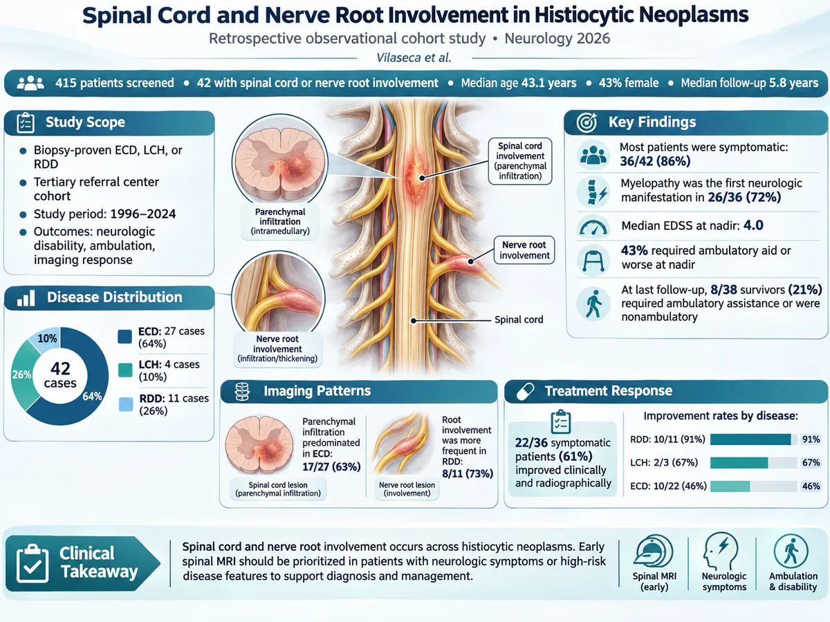
From a total of 415 patients (183 ECD, 144 LCH, 88 RDD), 42 exhibited spinal cord or nerve root involvement detectable on MRI (ECD 27, LCH 4, RDD 11). The median age at neurologic onset was 43.1 years, with a slight male predominance (57%). A large majority (86%) were symptomatic, presenting primarily with myelopathy (72%), which includes signs of spinal cord dysfunction such as weakness, sensory deficits, and sphincter disturbances.
Imaging Patterns: Parenchymal spinal cord infiltration was most common in ECD patients (63%) compared with LCH (50%) and RDD (18%), a statistically significant difference (p=0.035). Conversely, nerve root involvement dominated in RDD (73%) compared to ECD (26%) and LCH (25%) (p=0.021). These findings highlight distinct neuroanatomic predilections depending on histiocytic disease subtype, reflecting potentially differing pathobiology.
Neurologic Disability and Clinical Outcomes: At nadir, patients exhibited moderate disability (median EDSS 4.0), and 43% required ambulatory aids or were nonambulatory. Following treatment, 61% of symptomatic patients improved clinically and radiographically; improvements were more frequent in RDD (91%) and LCH (67%) than in ECD (46%) (p=0.022). At a median follow-up of 5.8 years, 21% of survivors remained severely disabled, requiring ambulatory assistance or being nonambulatory, emphasizing the potential for persistent morbidity.
Treatment Modalities: While detailed therapies were not exhaustively reported, the variable clinical responses suggest differing sensitivities to available treatments among the histiocytic subtypes. RDD and LCH appeared more amenable to therapy with improved neurologic outcomes compared to ECD, which often has a more insidious and refractory course.
Expert Commentary
This study is among the largest to systematically detail spinal cord and nerve root involvement in histiocytic neoplasms, filling a significant knowledge gap. Its findings underline the value of routine spinal MRI in patients with histiocytosis presenting with neurologic symptoms, facilitating earlier detection. The differential pattern of involvement—parenchymal infiltration in ECD and radicular involvement in RDD—may reflect differences in histiocyte distribution, inflammatory milieu, and molecular drivers, informing tailored diagnostic suspicion.
Despite its strengths, limitations include retrospective design, potential referral bias inherent to tertiary centers, and the relatively small number of LCH cases with spinal involvement limiting generalizability. Additionally, the study does not detail specific treatment regimens or molecular profiles, which are increasingly relevant given emerging targeted therapies informed by mutational status (e.g., BRAF V600E mutations in ECD and LCH).
Biologically, the preferential spinal parenchymal infiltration by ECD histiocytes might relate to their lipid-laden, inflammatory phenotype and proclivity for infiltrative fibrosis, contrasting with RDD cells which often cause prominent lymph node and nerve root swelling due to massive histiocytic proliferation and emperipolesis. Understanding these pathomechanisms may guide future therapeutic development and precision medicine approaches.
Conclusion
Spinal cord and nerve root involvement are important and clinically impactful manifestations of histiocytic neoplasms. Distinct radiologic and clinical patterns among ECD, LCH, and RDD warrant high clinical suspicion and support incorporating spinal MRI into diagnostic workups for patients with neurologic symptoms or high-risk disease. Early recognition and intervention can improve neurological outcomes and reduce long-term disability. Continued research into underlying mechanisms and targeted therapies holds promise for further improving patient care in these complex and heterogeneous disorders.
Funding and ClinicalTrials.gov
The original study was conducted with institutional support and involvement of the Histio-Care Network. No specific external funding sources or clinical trial registrations were reported.
References
Vilaseca A, Borre C, Go R, et al. Spinal Cord and Nerve Root Involvement in Histiocytic Neoplasms: A Retrospective Observational Cohort Study. Neurology. 2026 Oct 15;107(9):e218559. PMID: 42879725.
Other references include literature on the pathobiology and treatment of ECD, LCH, and RDD, typical nervous system involvement patterns, and the role of imaging in histiocytic disorders.
Specialty calculators
For clinical reference; use professional judgment.