

Highlight
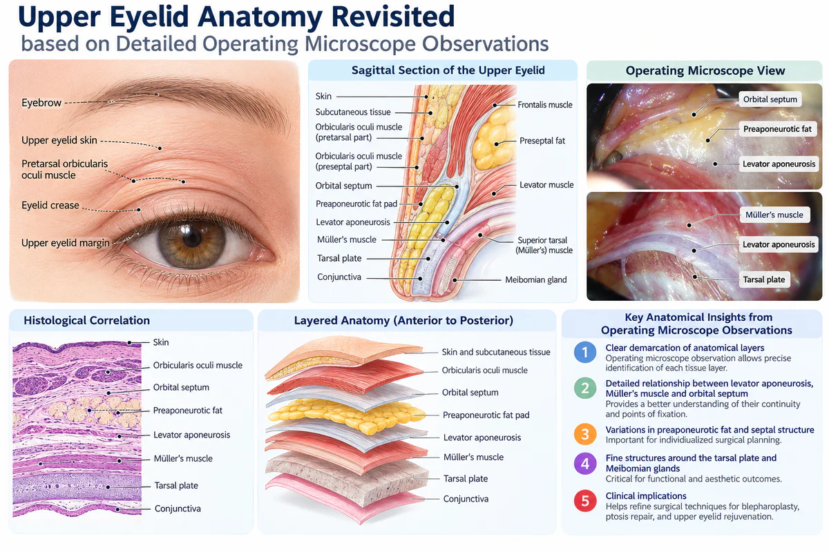
- Operating microscope analysis of 148 upper eyelids provides enhanced visualization of key upper eyelid structures including the orbital septum, retro-orbicularis oculi fat (ROOF), and levator aponeurosis.
- The study identifies the lower-positioned transverse ligament (LPTL) in 94% of eyelids and describes its anatomical relationships and clinical significance.
- Detailed measurements of the levator aponeurosis length and its bilaminar structure clarify its surgical relevance in upper blepharoplasty.
- An anterior surgical approach to the Müller muscle and the identification of critical bleeding danger zones improve operative safety and outcomes.
Study Background
Upper eyelid blepharoplasty is a commonly performed cosmetic and reconstructive procedure aimed at correcting eyelid ptosis, dermatochalasis, and restoring youthful upper eyelid contour. Despite its frequency, variations and controversies in the detailed anatomy of the upper eyelid have persisted, occasionally contributing to inconsistent surgical outcomes and complication risks such as bleeding or eyelid dysfunction. Clarifying the intricate anatomic structure, particularly under magnification, can enhance precision in tissue dissection and manipulation, thus improving both safety and aesthetic results.
Study Design
This anatomical study was conducted through detailed intraoperative observations under an operating microscope during 74 consecutive primary upper blepharoplasty surgeries, encompassing 148 eyelids from 54 Asian and 20 Caucasian patients. The approach allowed for high-resolution visualization of eyelid layers and anatomical relationships. Measurements of key structures were made, and a systematic dissection approach was documented, including exposure of the Müller muscle by an anterior approach. The presence and characteristics of the lower-positioned transverse ligament were assessed, alongside identification of safe zones to minimize intraoperative bleeding.
Key Findings
Orbital Septum and Adjacent Structures: Enhanced microscope visualization revealed the orbital septum as a dynamic anatomical barrier closely associated with the retro-orbicularis oculi fat (ROOF), levator aponeurosis, and retroseptal fat pads. These layers’ spatial relationships were delineated with clarity, aiding precise dissection planes.
Lower-Positioned Transverse Ligament (LPTL): The LPTL was identified in 94% of upper eyelids examined, validating it as a consistently present structure. Its anatomical location was elucidated in relation to the anterior fascial sheath of the levator aponeurosis and the bilaminar nature of the levator aponeurosis. The levator aponeurosis consists of two distinct layers: an anterior lamina and a posterior lamina, with the proximal fused portion averaging 5.4 mm in length (range 4.0–8.0 mm), and the full length averaging 17.6 mm (range 14–21 mm).
Anatomical Implications for Surgical Technique: The study’s findings support an anterior approach to the Müller muscle allowing better visualization and safer dissection. Correlation of the LPTL and levator aponeurosis architecture assists surgeons in identifying key landmarks, thus avoiding inadvertent tissue damage or inadequate fat pad excision.
Bleeding Danger Zones: Operating microscope observation facilitated detailed mapping of vascular anatomical danger zones in the upper eyelid. The study emphasizes adopting surgical strategies informed by these observations to mitigate bleeding complications during blepharoplasty.
Expert Commentary
The comprehensive microscopic approach redefines our understanding of upper eyelid anatomy, providing a precise surgical roadmap. These findings align with prior anatomical studies while refining the accuracy of structural definitions, especially concerning the lower-positioned transverse ligament’s role and the bilaminar levator aponeurosis. Expert consensus increasingly advocates such refined anatomical knowledge to guide tailored, safer blepharoplasty techniques. However, future studies could expand ethnic diversity and evaluate functional outcomes correlated to these anatomical landmarks to strengthen generalizability.
Conclusion
This study offers a pivotal contribution to clinical anatomy and surgical technique in upper eyelid blepharoplasty through detailed operating microscope observations. Recognition of the lower-positioned transverse ligament’s prevalence, levator aponeurosis bilaminar structure, and danger zones provides critical insights for surgeons. Incorporating these anatomical nuances into practice can enhance operative safety, outcome predictability, and overall patient satisfaction in both cosmetic and reconstructive eyelid surgery.
Funding and ClinicalTrials.gov
The published article does not specify funding sources or clinical trial registration information.
References
Wong CH, Hsieh MKH, Goy TWT, Minelli L, Mendelson B. Upper Eyelid Anatomy Revisited based on Detailed Operating Microscope Observations. Plast Reconstr Surg. 2026 Oct 21. PMID: 42904892. Available from: https://pubmed.ncbi.nlm.nih.gov/42904892/
Selva D, Chen CB. Anatomy and surgical considerations of blepharoplasty. Curr Opin Ophthalmol. 2012 Mar;23(2):122-7. doi:10.1097/ICU.0b013e32835089ca.
Obagi S, Davis D. Comprehensive understanding of upper eyelid anatomy in blepharoplasty: implications for aesthetic outcomes. Aesthet Surg J. 2018;38(6):635-647.