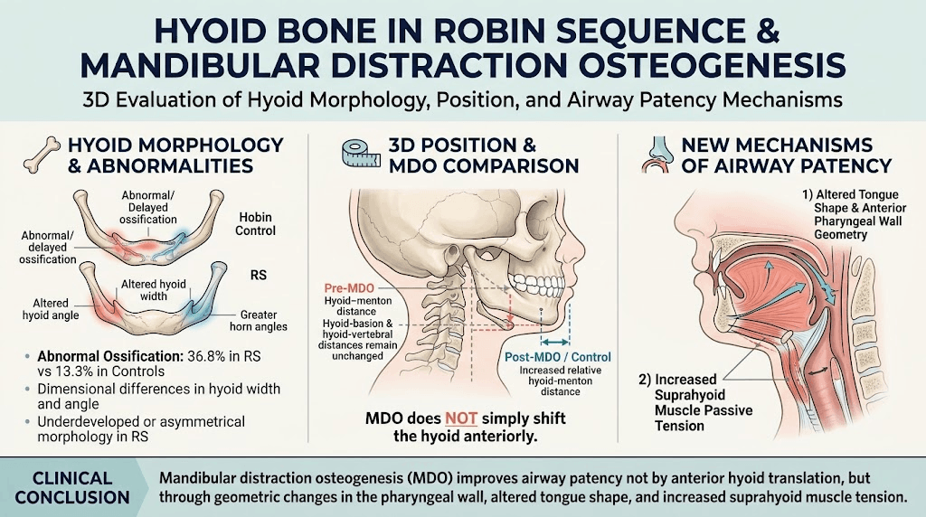
Patients with Robin Sequence (RS) exhibit distinct hyoid bone morphology and positional differences compared to controls.
Mandibular distraction osteogenesis (MDO) does not significantly move the hyoid bone anteriorly as previously hypothesized.
MDO likely improves airway patency by altering tongue shape, pharyngeal wall geometry, and increasing tension in suprahyoid muscles.
The study emphasizes the hyoid bone and its muscular attachments as key contributors to airway obstruction in RS.
Study Background
Robin Sequence (RS) is a congenital craniofacial condition characterized predominantly by micrognathia (small lower jaw), glossoptosis (downward displacement or retraction of the tongue), and subsequent upper airway obstruction. These anatomical abnormalities contribute significantly to feeding difficulties, respiratory distress, and may cause life-threatening airway compromise in neonates. The hyoid bone, a U-shaped bone in the anterior neck, serves as an anchoring structure for muscles involved in swallowing and respiration. In RS, the morphology and position of the hyoid bone are altered, influencing airway dimensions.
Mandibular distraction osteogenesis (MDO) has emerged as a surgical intervention in severe RS cases to lengthen the mandible, which is hypothesized to open the oropharyngeal airway by advancing the tongue and associated structures, including the hyoid bone. However, the exact role of hyoid positioning in airway improvement post-MDO remains unclear. This study aims to quantitatively analyze three-dimensional (3D) hyoid bone position and morphological characteristics in RS patients before and after MDO, compared to age-matched pediatric controls, to elucidate mechanisms underlying airway patency improvements.
Study Design
This retrospective observational study utilized computed tomography (CT) imaging data from 38 pediatric patients diagnosed with RS who underwent MDO and 15 control patients without RS. The RS group was evaluated pre- and post-MDO using CT scans to determine hyoid bone position relative to craniomandibular landmarks, including distances to the menton (chin), basion (base of skull), vertebral column, and temporomandibular joint (TMJ). Hyoid bone morphology parameters—such as width and angular measurements—were also assessed. Comparisons were made between pre-MDO, post-MDO, and control groups to identify significant positional and morphological differences.
Key Findings
Pre-MDO RS patients demonstrated significantly shorter relative distances from the hyoid bone to the menton compared to both post-MDO RS patients and controls (p<0.01). This suggests preoperative hyoid bone is positioned more posteriorly or inferiorly relative to the chin in RS. In contrast, the relative distances from the hyoid to the basion and vertebral column did not differ significantly among groups, indicating stable vertical and sagittal positioning relative to the skull base and cervical spine.
Notably, control patients exhibited smaller hyoid-temporomandibular joint distances compared to RS patients (p<0.05), implying lateral displacement or altered relations of the hyoid relative to TMJ in RS.
Morphologically, RS patients showed increased prevalence of abnormal hyoid ossification patterns (36.8% pre-MDO) compared to controls (13.3%). Additionally, dimensional analyses revealed RS-related differences in hyoid width and angular configuration, which changed further following MDO intervention.
Importantly, MDO did not produce the expected anterior displacement of the hyoid bone, challenging the hypothesis that airway enlargement post-MDO directly results from hyoid forward movement. Alternative mechanisms suggested include 1) alterations in tongue shape and anterior pharyngeal wall geometry induced by mandibular advancement and 2) increased passive tension in suprahyoid muscles anchored to the hyoid, which contribute to airway stabilization.
Expert Commentary
The findings provide important mechanistic insights into airway management in RS. The lack of anterior hyoid migration post-MDO suggests that airway patency improvements are mediated by complex soft tissue and muscular dynamics rather than skeletal repositioning alone. This underscores the pivotal role of the suprahyoid musculature and tongue repositioning in restoring airway function. Furthermore, the observed abnormal ossification and morphological changes in the hyoid bone point to intrinsic developmental variations in RS that may affect surgical planning and outcomes.
Limitations of the study include retrospective design and CT imaging dependency, which may not fully capture dynamic functional changes during respiration and swallowing. Future studies incorporating dynamic imaging and muscle function assessments alongside skeletal analysis would enhance understanding. Nevertheless, these findings are consistent with emerging literature highlighting the multidisciplinary components of airway obstruction in RS beyond mandibular hypoplasia.
Conclusion
Mandibular distraction osteogenesis in Robin Sequence does not primarily facilitate airway enlargement through anterior displacement of the hyoid bone. Instead, the improvement in airway patency appears to stem from modified tongue morphology, altered anterior pharyngeal wall configuration, and increased tension in muscles attaching to the hyoid. Recognition of the key role of the hyoid bone and its soft tissue connections expands the clinical understanding of RS pathophysiology and may inform enhanced surgical strategies. Further research exploring functional muscle dynamics and long-term morphological changes is warranted to optimize treatment outcomes.
Funding and Clinical Trial Registration
The original article does not specify sources of funding or clinical trial registration details.
References
1. Wang AT, Nathan SL, Prescher H, Ross CF, Reid RR, Li P. The Hyoid Bone in Robin Sequence: Morphology and Position in the Context of Mandibular Distraction Osteogenesis. Plast Reconstr Surg. 2026 Oct 19. PMID: 42896307.
2. Sher AE. Robin sequence and upper airway obstruction. Otolaryngol Clin North Am. 2004 Apr;37(2):279-297.
3. Evans KN, Sie KC. The surgical management of upper airway obstruction in Robin sequence. Curr Opin Otolaryngol Head Neck Surg. 2009 Dec;17(6):450-454.
4. Moura PH, et al. Evaluation of hyoid bone position among obstructive sleep apnea syndrome patients. Braz J Otorhinolaryngol. 2015 Jan-Feb;81(1):32-7.
5. Douglas JE, et al. Functional anatomy of the hyoid and suprahyoid muscles for pharyngeal airway stability. J Biomech. 2018 Nov;79:102-109.
6. Hunter JV, et al. Mandibular distraction osteogenesis: Imaging findings and complications. Radiographics. 2007 Jul-Aug;27(4): 1283-1300.