Highlights
- Quantitative computed tomography (CT) metrics of mucus plugs and emphysema define novel COPD subtypes beyond conventional clinical assessment.
- The mixed mucus plug-emphysema subtype presents the highest risk of acute exacerbations, demonstrating synergistic predictive value for clinical outcomes.
- Distinct COPD pathophysiological profiles identified by imaging correlate with exposure histories and have differential exacerbation risks and treatment responses.
- Incorporating quantitative imaging biomarkers enables refined personalized risk stratification and may guide targeted prevention strategies in COPD management.
Background
Chronic obstructive pulmonary disease (COPD) is a heterogeneous respiratory condition characterized by airflow limitation, emphysematous destruction, and airway pathology including inflammation and mucus hypersecretion. Traditional clinical tools and spirometry inadequately capture this heterogeneity, limiting prognostic accuracy and personalized therapeutic approaches. Acute exacerbations of COPD (AECOPD) markedly increase morbidity, mortality, and healthcare utilization, yet predicting exacerbation risk remains challenging.
Advances in quantitative computed tomography (CT) have enabled objective assessment of emphysema severity (% low attenuation area, %LAA) and airway abnormalities, including mucus plugs. These imaging biomarkers offer promise for classifying COPD subtypes with distinct pathophysiology and clinical trajectories. Recent prospective evidence suggests that quantitative CT-derived mucus plug scores and emphysema quantification jointly identify subgroups at differential exacerbation risk, supporting their utility for improved phenotyping and management.
Key Content
Prospective Evidence for Mucus Plug and Emphysema Phenotyping in COPD
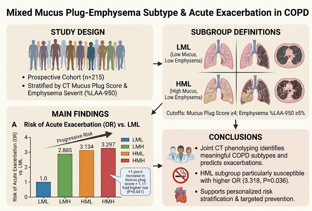
A landmark prospective cohort study by Li et al. (2026, Chest) investigated the prognostic significance of combined quantitative CT measures of mucus plugs and emphysema in 215 patients with COPD. Patients were stratified into four subgroups based on mucus plug score (cutoff: 4) and emphysema severity (%LAA-950 cutoff: 5%): low mucus/low emphysema, low mucus/high emphysema, high mucus/low emphysema, and high mucus/high emphysema.
Key findings included:
– Compared with the low mucus/low emphysema group, adjusted odds ratios (ORs) for acute exacerbation progressively increased across other groups: low mucus/high emphysema (OR 2.885), high mucus/low emphysema (OR 3.134), and high mucus/high emphysema (OR 3.297).
– Each 1-point increase in mucus plug score conferred a 1.1-fold increased exacerbation risk.
– The association of higher mucus plugs with exacerbations was significant predominantly in the low emphysema subgroup, underscoring distinct pathophysiological contributions.
This study substantiates the synergistic value of combined mucus plug and emphysema assessment as markers of COPD heterogeneity and predictors of exacerbation risk.
Comparative Phenotyping: Biomass vs Cigarette Smoke COPD Subtypes
A large real-world prospective observational study from the RealDTC cohort (PMID 42112598) compared COPD patients with biomass smoke exposure (BS-COPD), cigarette smoke exposure (CS-COPD), and combined exposures (CS+BS-COPD). Using quantitative CT, the study revealed:
– BS-COPD patients had significantly fewer emphysematous lesions (lower %LAA) but more airway mucus plugs and thicker airway walls.
– CS+BS-COPD patients exhibited the highest presence of both emphysema and mucus plugs.
– BS-COPD patients demonstrated lower risk of moderate-to-severe exacerbations compared to CS-COPD, but combined exposures yielded the highest exacerbation incidence.
– Inhaled therapy combinations including LABA+LAMA or ICS+LABA+LAMA were associated with reduced exacerbation risk versus LAMA monotherapy.
This evidence highlights distinct structural and functional phenotypes related to exposure history with differential exacerbation risks, supporting integration of quantitative CT phenotyping in clinical assessment.
Emerging Multidimensional Profiling for COPD Disease Stability
The J-UNMET-COPD study protocol (PMID 41881893) describes a prospective multicenter cohort aiming to integrate clinical phenotyping with quantitative imaging (emphysema %, airway wall thickness, mucus plugs), inflammatory biomarkers, and omics analyses to delineate phenotypes associated with disease stability and exacerbation risk.
This comprehensive approach exemplifies the next frontier in COPD research, moving beyond singular imaging biomarkers to multidimensional profiles that may predict treatment response and long-term outcomes, facilitating biomarker-guided precision medicine.
Expert Commentary
The integration of quantitative CT-derived mucus plugging and emphysema measures marks a significant advancement in COPD phenotyping. Mucus plugs reflect airway obstruction and inflammation-driven secretions, while emphysema represents parenchymal destruction; their combined presence delineates complex pathophysiologic subtypes that influence exacerbation risk.
Notably, the higher exacerbation risk in the mixed high mucus/high emphysema subgroup suggests additive or synergistic pathogenic mechanisms. The observation that mucus plugs independently predict exacerbations primarily in low emphysema patients indicates that airway-centric pathology can dominate clinical severity in the absence of extensive parenchymal damage.
Clinically, these findings advocate incorporating quantitative CT metrics into routine COPD evaluation to enable personalized risk stratification. Identifying patients with high mucus plugging may prompt intensified airway clearance strategies and tailored pharmacologic approaches, while those with predominant emphysema may benefit from alternative interventions.
However, limitations exist including accessibility of quantitative CT in routine practice, radiation exposure considerations, and the need for standardized scoring algorithms and prospective validation across diverse populations. Furthermore, the relationship between imaging phenotypes and molecular inflammatory profiles remains an active area of research with translational potential.
Guidelines have yet to formally endorse quantitative imaging for phenotyping, but accumulating evidence may shift paradigms toward imaging-assisted personalized COPD care.
Conclusion
Recent prospective evidence firmly establishes the prognostic value of combined quantitative CT-derived mucus plug scores and emphysema metrics in predicting acute COPD exacerbations, defining novel subtypes within the heterogeneous COPD spectrum. Complementary findings from population studies of smoke exposure subtypes underscore the distinct pathophysiology and exacerbation risks associated with structural and airway abnormalities.
These advances highlight the promise of imaging biomarkers to refine COPD phenotyping, enabling personalized risk stratification and targeted prevention strategies. Future directions include expanding multidimensional profiling integrating imaging, molecular biomarkers, and clinical data to optimize disease stability and guide precision therapeutics.
Clinicians and researchers should consider the emerging role of quantitative CT in COPD management, advocating for broader implementation, standardization, and integration within clinical trials and real-world practice.
References
- Li Y, Wang Y, Zhang M, et al. Mixed Mucus Plug-Emphysema Subtype and Acute Exacerbation in Chronic Obstructive Pulmonary Disease. Chest. 2026 Sep 19. PMID: 42762984. https://pubmed.ncbi.nlm.nih.gov/42762984/
- Liang J, Ma Y, Zhang P, et al. Clinical features and longitudinal assessment in outcomes of chronic obstructive pulmonary disease with biomass smoke exposure history: A prospective study of the RealDTC cohort. Pulmonology. 2026 Dec;32(1):2671536. doi:10.1080/25310429.2026.2671536. PMID: 42112598. https://pubmed.ncbi.nlm.nih.gov/42112598/
- Tanaka T, Nakamura H, Yamamoto M, et al. A prospective, multicenter cohort study to identify unmet medical needs in Japanese patients with chronic obstructive pulmonary disease: The J-UNMET-COPD study protocol. Respir Investig. 2026 May;64(3):101409. doi:10.1016/j.resinv.2026.101409. PMID: 41881893. https://pubmed.ncbi.nlm.nih.gov/41881893/