Highlight
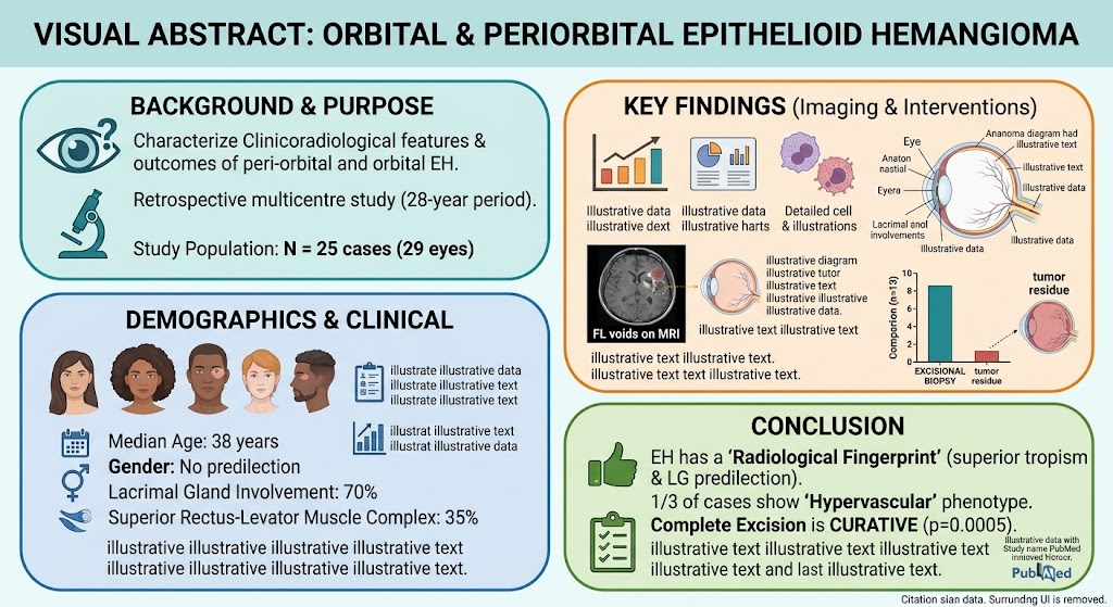
Orbital and periorbital epithelioid hemangioma (EH) predominantly affects the superior orbital quadrant with frequent lacrimal gland involvement. A distinct hypervascular radiological phenotype was identified in approximately one-third of cases on MRI and angiography. Complete surgical excision was significantly associated with curative outcomes, whereas infiltrative lesion patterns on imaging predicted tumor residue.
Study Background
Epithelioid hemangioma, also known as angiolymphoid hyperplasia with eosinophilia, is a rare benign vascular tumor that can occur in the orbital and periorbital regions. Despite being benign, the lesion’s infiltrative growth pattern, location near critical ocular structures, and potential for recurrence pose diagnostic and therapeutic challenges. Orbital EH has been under-characterized due to its rarity and disparate case reports. This study addresses the unmet need for a consolidated clinicoradiological understanding and evidence-based treatment approach through a multicentre retrospective series.
Study Design
This retrospective, multicentre, observational case series reviewed 25 biopsy-proven orbital and peri-orbital EH cases (29 affected eyes) managed across six tertiary referral centres over a 28-year period. The cohort was evaluated for clinical presentation, radiological characteristics from MRI and angiography, interventions, histopathological confirmation, and treatment outcomes, particularly tumor excision completeness and residual disease. Previously reported imaging features were pooled for comparative analysis.
Key Findings
Demographics and Clinical Presentation: The median patient age at diagnosis was 38 years (interquartile range 18–49 years), with no significant gender predisposition. Most cases presented with orbital lesions rather than solely periorbital involvement.
Radiological Patterns: Of 21 cases with imaging available, 80% (20 eyes) had orbital involvement. The superior orbital quadrant was the predominant site (70%). The lacrimal gland (LG) was involved in 70% of eyes, underscoring its susceptibility. Extraocular muscle involvement was noted in half of the eyes, especially the superior rectus-levator muscle complex (35%).
A notable subset (36%) exhibited a ‘‘hypervascular’’ phenotype on MRI characterized by heterogeneous contrast enhancement with internal flow voids, correlating with an intrinsic vascular blush on angiography. This imaging fingerprint can aid in differential diagnosis from other orbital masses.
Treatment Modalities and Outcomes: Interventions included excisional biopsy (62%), debulking (14%), incisional biopsy (24%), and primary sclerotherapy (5%). Complete excisional biopsy was statistically associated with curative outcomes (p=0.0005). Tumor residue was significantly associated with ill-defined, infiltrative lesions on imaging (p=0.0196), suggesting the critical value of thorough preoperative radiological assessment.
Comparison with Literature: The study pooled previous radiological descriptions and confirmed a consistent pattern of superior orbital tropism and vascular characteristics that distinguish EH from other orbital vascular lesions.
Expert Commentary
EH remains a diagnostic challenge due to its rarity and clinical overlap with other vascular and inflammatory orbital conditions. This study substantiates the importance of advanced imaging to detect hypervascular phenotypes and evaluate lesion margins, facilitating surgical planning. Complete excision, while sometimes technically challenging due to local infiltration, offers the best chance of cure, highlighting the necessity for experienced orbital surgeons and multidisciplinary care teams.
Limitations include the retrospective nature and heterogeneous imaging modalities over a long period, yet the multicentre design enhances generalizability. Prospective studies incorporating standardized imaging and long-term follow-up would further clarify recurrence rates and refine management strategies.
Conclusion
Orbital and periorbital epithelioid hemangioma exhibits distinctive clinicoradiological features, including superior orbital location, lacrimal gland and extraocular muscle involvement, and a hypervascular MRI phenotype in a subset of cases. Complete surgical excision, when feasible, is curative and should be the preferred treatment approach. Radiological evaluation is paramount for identifying lesion extent and potential infiltration to optimize surgical outcomes. This study advances the understanding of EH’s diagnostic and therapeutic nuances, guiding clinicians in managing this rare orbital tumor effectively.
Funding and ClinicalTrials.gov
No specific funding sources or clinical trial registrations were reported for this study.
References
1. Agarwal A, Almater A, Raghunath P, et al. Orbital and Periorbital Epithelioid Hemangioma (Angiolymphoid Hyperplasia with Eosinophilia): A multicentre study of 25 cases. Am J Ophthalmol. 2026 Sep 27;174(3):1-12. PMID: 42801951.
2. Shields JA, Shields CL. Tumors of the conjunctiva and cornea. Indian J Ophthalmol. 2017;65(5):393-400.
3. Rootman J. Diseases of the orbit: a multidisciplinary approach. 2nd ed. Philadelphia: Lippincott Williams & Wilkins; 2003.
4. Ginat DT, Vuong B. Orbital vascular lesions: radiological review. Radiol Clin North Am. 2014;52(1):63-78.