Highlight
- This study analyzes differences between radiographic and pathology-integrated metrics in assessing treatment response to neoadjuvant imatinib in KIT exon 11-mutant GIST.
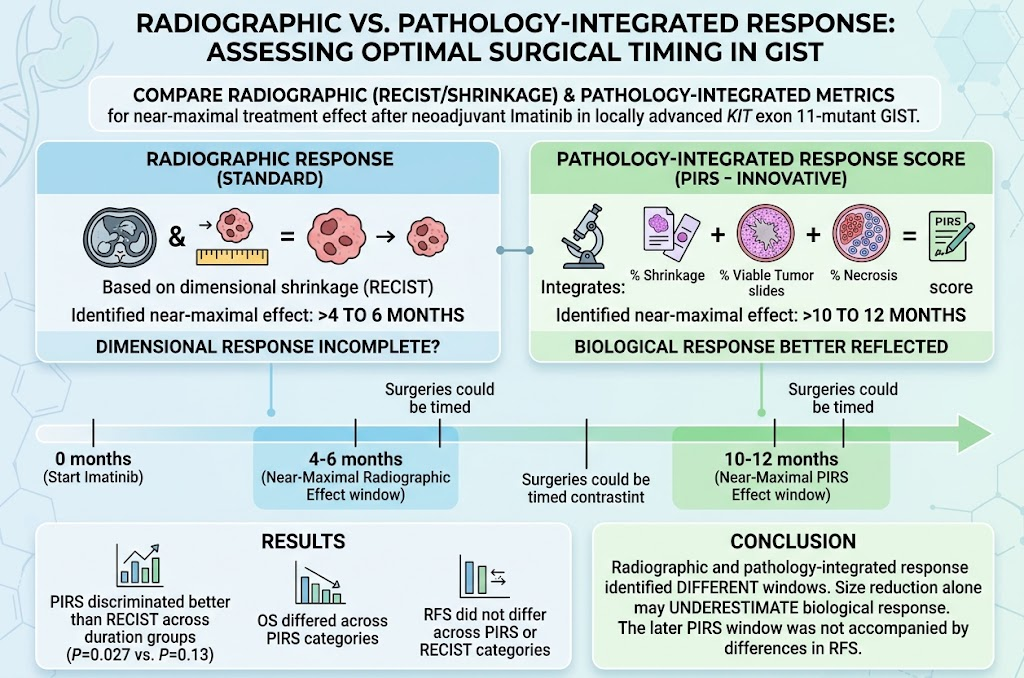
- Radiographic responses plateau earlier (4–6 months), whereas pathology-integrated response peaks later (10–12 months), revealing a delayed maximal biological effect beyond size reduction.
- Pathology-integrated response better discriminated outcomes, including overall survival differences, compared to radiographic criteria like RECIST.
- Stable disease by RECIST did not always indicate poor pathology response, underscoring the limitations of size-based imaging alone in guiding surgical timing.
Study Background
Gastrointestinal stromal tumors (GIST) harboring mutations in the KIT exon 11 gene represent a common molecular subtype with sensitivity to targeted therapy via imatinib mesylate. Neoadjuvant imatinib is increasingly utilized to downstage locally advanced GIST, facilitating potentially curative surgical resection. Determining the optimal timing for surgery remains a clinical challenge. Traditionally, radiographic shrinkage assessed by Response Evaluation Criteria in Solid Tumors (RECIST) guides surgical timing. However, dimensional changes may not fully capture the biological treatment effect, such as viable tumor cell death and necrosis, which are crucial for long-term prognosis. This gap creates uncertainty whether radiographic criteria alone suffice to define maximal therapeutic response and the best surgical window.
Study Design and Methods
This retrospective cohort study included 131 patients with locally advanced KIT exon 11-mutant GIST treated between 2011 and 2022 at two major sarcoma centers in the United States and Italy. All patients received neoadjuvant imatinib followed by curative-intent resection. Radiographic response was assessed via RECIST and percentage tumor shrinkage at two-month intervals for a period ranging from 3 to 22 months. Importantly, the investigators introduced a pathology-integrated response score (PIRS) evaluating composite metrics: tumor size shrinkage, percentage of viable tumor cells, and degree of necrosis from surgical specimens. This allowed correlation of radiographic changes with pathological evidence of treatment effect. The primary endpoint was identification of the earliest time interval when patients achieved 90% of the peak median response according to each metric.
Key Findings
The median patient age was 62 years; 60.3% were male. Nearly half achieved partial response by RECIST (45.8%), while 48.9% had stable disease during neoadjuvant treatment. Radiographic assessment indicated that near-maximal tumor shrinkage was reached between 4 to 6 months after treatment initiation. Conversely, PIRS showed the near-maximal pathological treatment effect emerged later, between 10 to 12 months.
Statistical analysis demonstrated that PIRS better distinguished patient groups based on treatment duration than RECIST (P=0.027 vs. P=0.13), indicating greater sensitivity of pathology measures in reflecting cumulative treatment effect.
Significantly, 30.5% of patients categorized as stable disease by RECIST had major or near-complete pathologic response, revealing that size criteria alone may underestimate tumor biology changes. Regarding clinical outcomes, overall survival varied significantly across PIRS-defined response categories (better survival with major pathological response), but recurrence-free survival did not differ by PIRS or RECIST status.
Expert Commentary
The study by Ranjbarian et al. offers critical insights into the timing of surgery post-neoadjuvant imatinib in KIT exon 11-mutant GIST. Traditional reliance on radiographic criteria alone appears insufficient to capture the full therapeutic impact, as tumor necrosis and viable cell reduction continue beyond the plateau of size shrinkage.
This delayed maximal pathological treatment effect may reflect ongoing tumor microenvironment remodeling that is not evident on imaging. Consequently, premature surgery based solely on imaging could truncate potential benefits of prolonged therapy, although the lack of difference in recurrence-free survival suggests a complex interplay requiring further investigation.
Notably, the study is limited by its retrospective design and potential heterogeneity in imaging intervals and surgical decision-making. Validation in prospective trials and integration of functional imaging or biomarker assessment might refine treatment response assessment.
Guidelines currently emphasize radiographic criteria for surgical timing, but these findings advocate for incorporating pathological and molecular endpoints to optimize clinical decision-making and personalize patient management.
Conclusion
This multi-institutional retrospective analysis delineates a significant divergence between radiographic and pathology-integrated response timelines in KIT exon 11-mutant GIST patients treated with neoadjuvant imatinib. While imaging-defined tumor shrinkage occurs earlier, the maximal pathological treatment effect—reflected by viable tumor cell death and necrosis—manifests later, suggesting that surgery may be optimally timed after achieving near-maximal pathology response rather than solely based on size reduction.
The findings underscore the importance of incorporating comprehensive pathological assessment into treatment evaluation to better tailor surgical timing and improve overall survival outcomes. Future prospective studies are warranted to validate these observations and assess how integrating pathology and advanced imaging biomarkers can refine the management of locally advanced GIST.
Funding and Clinical Trials
The original study did not specify external funding sources. The analysis was conducted across two tertiary sarcoma centers with no declared conflicts of interest. No clinical trials registration was indicated for this retrospective cohort.
References
1. Ranjbarian T, Del Simone M, Kang Sim DE, et al. Radiographic Versus Pathology-Integrated Response for Assessing Optimal Surgical Timing After Neoadjuvant Imatinib in Patients With Locally Advanced KIT Exon 11-Mutant GIST. Ann Surg. 2026 Jun 26;284(3):506-515. PMID: 42440135.
2. Demetri GD, von Mehren M, Antonescu CR, et al. NCCN Guidelines Insights: Soft Tissue Sarcoma, Version 2.2018. J Natl Compr Canc Netw. 2018;16(5):536-563.
3. Eisenhauer EA, Therasse P, Bogaerts J, et al. New response evaluation criteria in solid tumours: revised RECIST guideline (version 1.1). Eur J Cancer. 2009;45(2):228-247.
4. Joensuu H. Gastrointestinal stromal tumor (GIST). Ann Oncol. 2006;17 Suppl 10:x280-x286.
5. Fletcher CD, Berman JJ, Corless C, et al. Diagnosis of gastrointestinal stromal tumors: a consensus approach. Hum Pathol. 2002;33(5):459-465.
6. Agaram NP, Wong GC, Guo T, et al. Pathologic Response to Imatinib in Gastrointestinal Stromal Tumors. J Gastrointest Surg. 2011;15(12):2080-2089.