Highlight
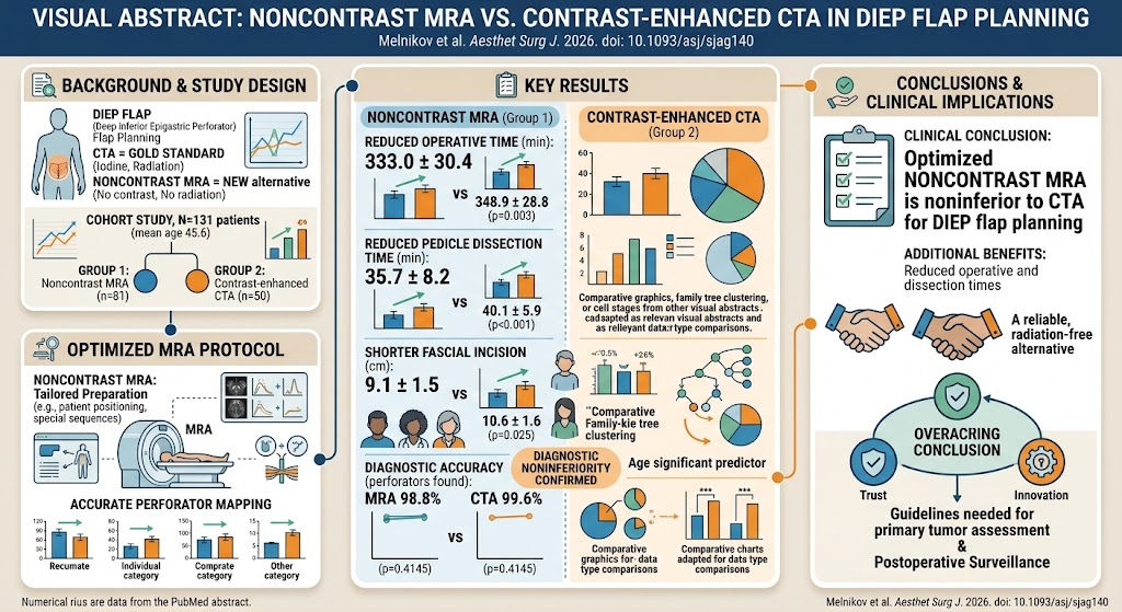
This study investigates an optimized noncontrast magnetic resonance angiography (MRA) technique for use in deep inferior epigastric perforator (DIEP) flap planning for breast reconstruction, challenging the conventional reliance on contrast-enhanced computed tomography angiography (CTA).
Key findings include noninferiority of noncontrast MRA compared to contrast-enhanced CTA based on diagnostic accuracy, with additional benefits of reduced operative and pedicle dissection times, shorter fascial incisions, and avoidance of ionizing radiation and iodinated contrast toxicity.
The comparable safety profile and imaging efficacy position noncontrast MRA as a promising, radiation-free alternative with clinical implications for preoperative vascular mapping in DIEP flap breast reconstruction.
Background
Deep inferior epigastric perforator (DIEP) flap breast reconstruction is a sophisticated surgical technique following mastectomy, capitalizing on the patient’s abdominal perforator vessels to create a natural, autologous breast mound. Preoperative vascular imaging is crucial to identify optimal perforators, minimize complications, reduce operative time, and improve surgical outcomes.
Contrast-enhanced computed tomography angiography (CTA) currently serves as the “gold standard” imaging modality due to its high spatial resolution and robust perforator visualization. However, CTA involves exposure to ionizing radiation and administration of iodine-based contrast agents that carry risks of nephrotoxicity and allergic reactions.
Magnetic resonance angiography (MRA) provides a radiation-free technique and, traditionally, gadolinium-based contrast is used to improve visualization. Nevertheless, concerns about gadolinium deposition, nephrogenic systemic fibrosis, and costs limit its routine use. Therefore, developing optimized noncontrast MRA protocols capable of accurate perforator mapping without contrast agents represents an unmet clinical need.
Study Design
This investigation was designed as a prospective, noninferiority cohort study over four years, involving 131 women undergoing delayed unilateral DIEP flap breast reconstruction. Inclusion criteria emphasized typical patient demographics (mean age ~45.6 years, BMI ~28.5 kg/m2) without contraindications to either imaging modality.
Participants were split into two groups: 81 women received an optimized noncontrast MRA with tailored preprocedural preparation and specialized scanning protocols to enhance imaging quality, while 50 women underwent standard contrast-enhanced CTA as per institutional protocol.
The primary endpoint was diagnostic accuracy of perforator mapping, with secondary endpoints including operative time, pedicle dissection time, fascial incision length, localization accuracy, and postoperative complication rates.
Key Findings
The study demonstrated that noncontrast MRA was noninferior to contrast-enhanced CTA in deep inferior epigastric perforator flap planning with a diagnostic accuracy of 98.8% versus 99.6% respectively (p=0.4145). Sensitivity and positive predictive values were similarly high, indicating excellent perforator visualization without contrast administration.
Notably, key surgical metrics favored the MRA group: total operative time decreased significantly (333.0 ± 30.4 minutes for MRA vs. 348.9 ± 28.8 minutes for CTA; p=0.003), as did pedicle dissection time (35.7 ± 8.2 vs. 40.1 ± 5.9 minutes; p<0.001). The mean length of fascial incision was shorter with MRA (9.1 ± 1.5 cm vs. 10.6 ± 1.6 cm; p=0.025), reflecting more precise preoperative mapping and potentially less surgical trauma.
The positional accuracy of perforator localization between modalities was comparable: the mean localization difference was 7.38 ± 2.59 mm for MRA versus 6.96 ± 2.34 mm for CTA (p=0.238), supportive of clinical interchangeability.
Importantly, no differences in perfusion-related complications occurred between groups, confirming that clinical safety and flap viability were maintained.
Expert Commentary
This study addresses a pivotal concern in oncoplastic reconstructive surgery—avoiding the cumulative radiation dose and contrast-induced nephropathy, especially in predominantly younger breast cancer survivors advocating for autologous reconstruction.
The successful application of an optimized noncontrast MRA protocol challenges entrenched reliance on CTA, aligning with trends prioritizing patient safety and resource stewardship without compromising surgical success.
However, potential caveats include the need for specialized MRA protocols and technician expertise, which might limit widespread adoption initially. Further, while the cohort size is reasonable, larger multicenter studies or randomized controlled trials could solidify evidence and ensure generalizability across diverse populations and imaging platforms.
Conclusion
This 4-year cohort study robustly supports optimized noncontrast magnetic resonance angiography as a clinically reliable, noninferior imaging technique for DIEP flap planning compared to contrast-enhanced CTA. It offers clear advantages by eliminating radiation exposure and iodinated contrast risks while also improving operative efficiency.
Adoption of noncontrast MRA protocols could enhance patient safety both preoperatively and longitudinally, making it a compelling alternative in reconstructive breast surgery.
Future research should focus on standardizing MRA protocols, assessing cost-effectiveness, and integrating advanced imaging analytics to further refine preoperative perforator mapping.
Funding and ClinicalTrials.gov
No specific funding information was provided in the article. The study registration or clinical trials information was not mentioned.
References
Melnikov DV, Abdeeva EI, Cotofana S, Ivanov SI, Ratushniy NA, Vorontsov AV, Gombolevskiy VA. Noncontrast Magnetic Resonance Angiography vs Contrast-Enhanced Computed Tomography in Deep Inferior Epigastric Perforator Flap Planning: A 4-year Noninferiority Cohort Study. Aesthetic surgery journal. 2026 Sep 16;46(10):1137-1146. PMID: 42413036. https://pubmed.ncbi.nlm.nih.gov/42413036/
Rozen WM, Whitaker IS, Stella DL, et al. Perforator mapping in DIEP flaps: the utility of preoperative imaging. Journal of Plastic, Reconstructive & Aesthetic Surgery. 2010;63(12):2049-2053.
Munhoz AM, Montag E, Paulista PL, et al. Deep inferior epigastric perforator flap breast reconstruction and imaging methods for preoperative perforator identification: a review of current concepts. Microsurgery. 2018;38(5):504-514.