
Highlight
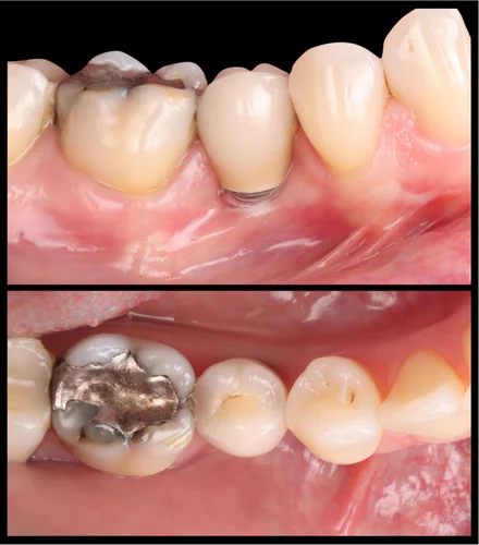
This 25-year prospective cohort study assessed the role of keratinized mucosa width (KMW) and mucosal thickness (MT) around tissue-level dental implants in partially edentulous patients with consistent oral hygiene and supportive care. Key findings include: (1) absence of KMW correlates with increased crestal bone loss and higher risk of peri-implant soft-tissue dehiscence; (2) mucosal thickness less than 2 mm is associated with increased soft-tissue complications but not with peri-implant disease or crestal bone loss; (3) neither KMW nor MT significantly influenced long-term brushing discomfort; (4) peri-implantitis prevalence was low and not significantly related to KMW or MT categories.
Study Background
Dental implants have become a cornerstone of rehabilitation in partially edentulous patients, offering functional and esthetic benefits. The long-term success of implants depends not only on osseointegration but also on the maintenance of healthy peri-implant soft tissues. Keratinized mucosa, the dense, tough gingiva surrounding teeth and implants, and mucosal thickness are hypothesized to influence peri-implant tissue health by providing a protective barrier against mechanical trauma and microbial invasion. However, the clinical significance of KMW and MT in preventing peri-implant diseases and ensuring patient comfort remains controversial. Addressing this gap, the current study provides important longitudinal data on how these mucosal parameters impact implant outcomes over an unprecedented 25-year follow-up period under consistent supportive peri-implant care and oral hygiene.
Study Design
This was a long-term observational cohort study including 149 partially edentulous patients rehabilitated with tissue-level implants. A total of 235 implants were analyzed with a mean follow-up duration of 25.4 7 years. Patients were enrolled based on adequate oral hygiene and adherence to regular supportive peri-implant maintenance. Clinical and radiographical evaluations were conducted by calibrated examiners. Data recorded included peri-implant probing depth (PD), bleeding on probing (BOP), suppuration on probing (SOP), plaque index, crestal bone loss (CBL), peri-implantitis prevalence, soft tissue dehiscence, and implant platform visibility. KMW was categorized into three groups: 0 mm (absent), <2 mm, and 2 mm. Mucosal thickness was classified as thin (<2 mm) and thick (≥2 mm). Patient-reported brushing discomfort was also recorded and analyzed in relation to mucosal parameters.
Key Findings
Peri-Implant Disease and Bone Stability
The prevalence of peri-implantitis was relatively low across groups: 11.1% at 0 mm KMW, 0% at <2 mm KMW, and 7.1% at ≥2 mm KMW, with no statistically significant differences (p=0.98). This suggests peri-implantitis occurrence may not directly depend on KMW when patients maintain proper oral hygiene and supportive care.
However, significant differences were observed in crestal bone loss. Implants with absent KMW showed greater bone loss compared to groups with KMW <2 mm (-0.28 mm difference) and ≥2 mm (-0.40 mm difference), with p=0.01. This highlights the protective role of keratinized mucosa in minimizing crestal bone resorption.
Soft Tissue Outcomes
The absence of KMW was associated with an increased risk of peri-implant soft-tissue dehiscence (PSTD) and visible implant platform exposure (p ≤ 0.01). Similarly, thin mucosal thickness was linked to increased PSTD (p=0.005) and implant platform visibility (p=0.04). Interestingly, thin mucosa was associated with a lower plaque index (p=0.01), possibly reflecting patient or site-specific factors.
Inflammatory Parameters and Clinical Measures
No significant differences related to KMW or MT were observed in probing depth, bleeding on probing, plaque index (except as noted above), or in the prevalence of peri-implant diseases (p > 0.05). Suppuration on probing, an indicator of active infection, was also not associated with mucosal parameters.
Patient-Reported Brushing Discomfort
Brushing discomfort was reported in 11.4% of sites with absent KMW, compared to 3.4% and 2.4% in the <2 mm and ≥2 mm groups, respectively. However, these differences did not achieve statistical significance (p>0.05). Similarly, mucosal thickness did not significantly influence brushing discomfort. This suggests that while mucosal parameters influence clinical tissue stability, their impact on patient-perceived discomfort during oral hygiene may be limited under well-maintained conditions.
Expert Commentary
This landmark study provides robust, long-term evidence for the role of keratinized mucosa in peri-implant tissue stability. The data underscore that adequate KMW can mitigate crestal bone loss and reduce risks of soft-tissue dehiscence and implant platform exposure. These findings align with biological plausibility, as keratinized mucosa likely offers mechanical protection against trauma and facilitates plaque control. The observation that mucosal thickness relates mainly to soft tissue esthetic complications, rather than inflammation or bone loss, refines our understanding of tissue phenotype influences.
The study’s strengths include its extensive follow-up, meticulous patient monitoring, and calibrated assessments. Limitations include the observational design and potential confounders related to patient-specific factors not fully controlled in the analysis. Furthermore, the patient cohort had excellent oral hygiene and frequent supportive care visits, which may limit generalizability to broader populations with variable compliance.
Current consensus documents and clinical guidelines emphasize the importance of peri-implant soft tissue management, but the optimal thresholds for KMW and MT remain debated. This study provides compelling data supporting a clinical preference for maintaining at least minimal keratinized mucosa around implants to ensure long-term stability.
Conclusion
In conclusion, this 25-year cohort study highlights the critical role of keratinized mucosa width in maintaining long-term peri-implant crestal bone stability and preventing soft tissue complications. While mucosal thickness influences esthetic soft tissue outcomes, neither KMW nor MT significantly impacts patient-reported brushing discomfort when oral hygiene is well maintained. These findings emphasize the importance of preserving or augmenting keratinized mucosa during implant placement and restorative procedures to optimize implant longevity and tissue health under supportive care. Further research may investigate strategies to enhance mucosal parameters in diverse patient populations and explore their impact on implant outcomes in less controlled clinical settings.
Funding and ClinicalTrials.gov
No specific funding sources or clinical trial registration were reported for this study.
References
- Couso-Queiruga E, Raabe C, Roccuzzo A, Salvi GE, Fonseca M, Chappuis V. Impact of Keratinized Mucosa Width and Mucosal Thickness on Implant Outcomes and Brushing Discomfort: A 25-Year Cohort Study. J Clin Periodontol. 2026 Aug;53(11):1691-1701. doi:10.1111/jcpe.14012. PMID: 42563453.
- Bouri A Jr, Bissada N, Al-Zahrani MS, Faddoul F, Nouneh I. Width of keratinized gingiva and the health status of the supporting tissues around dental implants. Int J Oral Maxillofac Implants. 2008 Jan-Feb;23(1):323-6.
- Schrott AR, Jimenez M, Hwang JW, Fiorellini JP, Weber HP. Clinical and histologic outcomes of sites with narrow vs. wide keratinized tissue. Int J Periodontics Restorative Dent. 2009 Oct;29(5):563-71.
- Bassi MA, Özcan M, Gherlone EF, et al. Influence of keratinized mucosa width on periimplant soft-tissue health and stability around dental implants: a systematic review and meta-analysis. Clin Oral Implants Res. 2021 Mar;32(3):317-332.
Specialty calculators
For clinical reference; use professional judgment.