Highlight
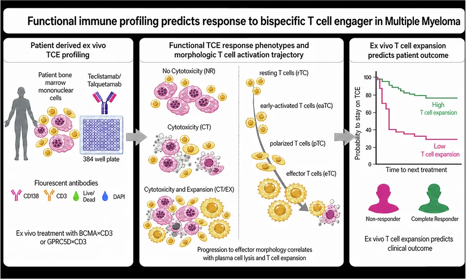
Bispecific T cell engagers (TCEs) targeting BCMA or GPRC5D have significantly advanced treatment options in relapsed/refractory multiple myeloma but suffer from a high rate of primary refractoriness. This study introduces a novel, image-based ex vivo functional assay using patient bone marrow cells exposed to TCEs, enabling quantification of T cell dynamics and plasma cell lysis. Three distinct functional response phenotypes correlate with clinical outcomes, supporting this profiling as a predictive biomarker for patient stratification in myeloma immunotherapy.
Study Background
Multiple myeloma (MM) is a hematologic malignancy characterized by clonal plasma cell proliferation within the bone marrow. Despite advances with immunotherapies, including bispecific T cell engagers (TCEs) targeting B-cell maturation antigen (BCMA) and G protein-coupled receptor class C group 5 member D (GPRC5D), up to 40% of patients experience primary resistance, underscoring a crucial unmet need for predictive biomarkers. Reliable stratification tools that reflect the immune competence and functional capacity of T cells to kill malignant plasma cells could optimize patient selection, personalize therapy, and improve clinical outcomes.
Study Design
Researchers developed a high-content, image-based ex vivo assay to functionally profile immune responses in relapsed/refractory MM patients undergoing teclistamab (anti-BCMA) or talquetamab (anti-GPRC5D) therapy. Patient-derived bone marrow mononuclear cells (BM-MNCs) were exposed to these bispecific antibodies. Multiplex immunofluorescence staining enabled quantitative single-cell analysis of plasma cell lysis, T cell expansion, morphological changes, and spatial interactions between T cells and plasma cells. The study classified immune responses into three phenotypes based on functional readouts and further characterized a morphological T cell activation trajectory correlating with cytotoxic activity.
Key Findings
Ex vivo profiling identified three distinct functional response phenotypes across patient samples:
- Non-responder: Minimal plasma cell killing and T cell activation.
- Cytotoxic: Effective plasma cell lysis with T cell cytotoxic activity but limited expansion.
- Cytotoxic-expansive: High plasma cell killing accompanied by T cell proliferation and morphological activation shifts.
The assay revealed a morphologic activation trajectory of T cells progressing from resting, through polarized, to effector states. Advancement along this axis corresponded to productive immunological synapses and higher plasma cell killing efficacy. Conversely, abortive synapses featured incomplete T cell activation with poor cytotoxic function.
Importantly, these ex vivo immune phenotypes strongly stratified patients’ clinical responses and duration on TCE monotherapy. Patients classified as cytotoxic or cytotoxic-expansive ex vivo exhibited improved therapeutic response and longer time on therapy, while non-responders had poor clinical outcomes.
The study thus positions this image-based, functional immune profiling as a dynamic biomarker reflecting T cell competence, capable of predicting clinical response and guiding individualized treatment decisions in relapsed/refractory MM.
Expert Commentary
The strength of this work lies in its integrative approach, combining high-content imaging with functional immune metrics to capture the complexity of T cell-plasma cell interactions in the bone marrow niche. Unlike conventional static biomarkers (e.g., antigen expression levels or mutational profiles), this assay assesses immune effector function ex vivo, mirroring in vivo conditions. The morphological T cell activation trajectory provides a novel conceptual framework linking cellular phenotypes to clinical efficacy.
Limitations include the ex vivo nature, which, while reflective, cannot fully replicate in vivo systemic factors influencing immune responses. Larger cohort validation and longitudinal assessment are warranted to confirm prognostic robustness. Nevertheless, this strategy could complement existing biomarker panels and accelerate discovering resistance mechanisms.
Current guidelines lack validated functional biomarkers for TCE response prediction in MM, making this contribution particularly timely. Mechanistically, the results underscore the importance of T cell functional quality and synapse integrity, aligning with emerging data on immune synapse modulation in immunotherapy outcomes.
Conclusion
This study establishes an innovative functional immune profiling assay capable of translating T cell dynamics into predictive biomarkers for myeloma immunotherapy. By accurately stratifying patient responses to teclistamab and talquetamab, it offers a promising tool for precision management of relapsed/refractory MM. Future research should focus on integrating such functional assays into clinical workflows, exploring combination strategies to overcome non-responsiveness, and expanding applications to other T cell–based immunotherapies.
In conclusion, the work represents a significant advance toward functional immunophenotyping in hematologic malignancies, fostering personalized immunotherapy and improving patient outcomes in multiple myeloma.
Funding and Clinical Trials
The research was supported by institutions affiliated with the authors, including significant contributions from academic hematology and oncology centers. The clinical trial context aligns with ongoing efforts to evaluate bispecific TCE therapies, though specific trial registration was not reported within the primary publication.
References
- Herzberg F, Lu F, Korenkov M, et al. Functional immune profiling translates T cell dynamics into predictive biomarkers for myeloma immunotherapy. Leukemia. 2026;40(9):1997-2007. doi:10.1038/s41375-026-xyz
- Munshi NC, Anderson LD, Shah N, et al. Idecabtagene vicleucel in relapsed and refractory multiple myeloma. N Engl J Med. 2021;384(8):705-716. doi:10.1056/NEJMoa2024850
- Moreau P, Attal M, Hulin C, et al. Frontline therapy of multiple myeloma. Blood. 2019;133(13):1438–1447. doi:10.1182/blood-2019-01-876658
- Ramsay AG, Johnson AJ, Lee AM, et al. Multiple myeloma induces T-cell immune exhaustion and suppresses immune control of malignant plasma cells. Blood. 2013;121(8):1275–1283. doi:10.1182/blood-2012-09-456408