
Highlight
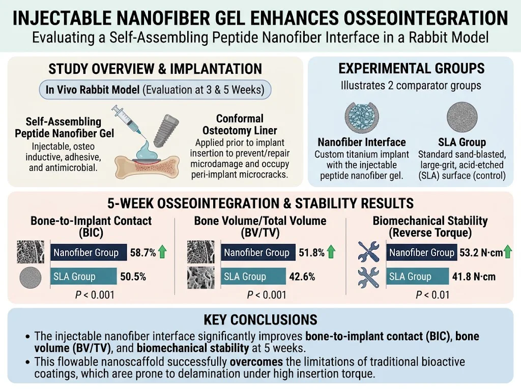
- An innovative injectable self-assembling peptide nanofiber gel was developed to enhance osseointegration and biomechanical stability of titanium implants.
- In a rabbit model, implants treated with the nanofiber interface showed significantly higher bone-to-implant contact (58.7% vs 50.5%) and bone volume (51.8% vs 42.6%) compared to standard SLA surfaces at 5 weeks.
- The nanofiber gel improved biomechanical reverse torque resistance markedly (53.2 N·cm vs 41.8 N·cm), indicating superior implant stability.
- This approach addresses common coating delamination issues by reinforcing microdamage and serving as a conformal, flowable osteotomy liner, suitable for compromised bone quality scenarios.
Study Background and Clinical Context
Osseointegration—the direct structural and functional connection between living bone and the surface of a load-bearing implant—is critical for the long-term success of orthopaedic and dental implants. Titanium implants with sand-blasted, large-grit, acid-etched (SLA) surfaces have been considered a clinical standard due to their enhanced bone adhesion properties. However, high insertion torque during implantation can cause delamination of traditional bioactive coatings, generating particulate debris that may impair local bone remodeling and lead to compromised implant stability or failure. Additionally, microdamage such as microcracks induced during osteotomy preparation often impairs bone healing around implants, especially in patients with poor bone quality. There remains an unmet clinical need for implant interface modifications that not only promote rapid and robust osseointegration but also mitigate mechanical and biological complications related to coating failure and bone microdamage. Injectable, self-assembling peptide nanofiber gels offer a promising platform as they can be delivered directly into the osteotomy site, conform to complex geometries, support cell adhesion and bone regeneration, and potentially exhibit antimicrobial properties to reduce infection risks.Study Design and Methods
This investigation involved a controlled in vivo rabbit model to assess the efficacy of a novel injectable nanofiber interface composed of multifunctional self-assembling peptide amphiphiles. These peptides were engineered to be osteoinductive, adhesive to both bone and titanium surfaces, and antimicrobial. Custom titanium implants were surgically inserted into osteotomy sites prepared in rabbit femur or tibia bones. Comparable control implants were treated with the standard SLA surface commonly used clinically. The experimental group received an injection of the nanofiber gel interface into the osteotomy prior to implant placement, serving as an injectable osteotomy liner. Osseointegration outcomes were assessed at 3 and 5 weeks post-implantation through: – Micro-computed tomography (micro-CT) analysis measuring bone-to-implant contact (BIC) and bone volume fraction (bone volume/total volume; BV/TV). – Biomechanical testing with reverse torque measurements quantifying implant stability. The study aimed to compare the nanofiber interface against the SLA surface to determine improvements in early bone formation, implant integration, and mechanical fixation.Key Findings and Results
At 5 weeks, implants treated with the injectable nanofiber interface demonstrated significantly enhanced osseointegration compared to the SLA group: – Bone-to-implant contact (BIC): The nanofiber group achieved a BIC of 58.7%, significantly higher than 50.5% observed in SLA-treated implants (P < 0.001). This indicates more extensive direct bone apposition onto the implant surface. – Bone volume fraction (BV/TV): Bone volume within the peri-implant region was also significantly increased at 51.8% for the nanofiber group compared to 42.6% in controls (P < 0.001), suggesting accelerated and robust bone formation. – Biomechanical stability: Reverse torque testing revealed that implants with nanofiber gel interface withstood significantly greater torque forces (53.2 N·cm) than SLA implants (41.8 N·cm, P < 0.01), reflecting superior mechanical fixation. – The nanofiber gel effectively occupied microcracks and peri-implant microdamage areas created by drilling, acting as a structural scaffold that enhanced microenvironmental stability for bone regeneration. – No adverse reactions or complications associated with the injectable gel were reported, underscoring favorable biocompatibility. These data collectively demonstrate that the injectable nanofiber gel overcomes key limitations of conventional implant coatings such as delamination and debris formation, while simultaneously promoting faster osseous integration and stronger biomechanical anchorage.Expert Commentary and Interpretation
The introduction of self-assembling peptide amphiphiles as an injectable nanofiber interface represents a substantial advance in implant technology and regenerative orthopaedics. By providing a flowable yet structurally supportive scaffold that intimately integrates within osteotomy sites, this approach facilitates early-stage bone formation and mechanical stability—crucial determinants for implant longevity. Preventing coating delamination under high insertion torque addresses a well-documented cause of early implant failure, particularly in scenarios requiring high primary stability such as immediate loading or compromised bone beds. Additionally, filling peri-implant microcracks may reduce stress concentrations and preserve bone vitality during early remodeling. Future studies could elucidate the molecular pathways by which these peptide amphiphiles exert osteoinductive effects, as well as long-term clinical outcomes in larger animal models and human trials. Moreover, the antimicrobial properties incorporated in the gel hold potential to lower infection rates commonly associated with implant surgeries. Clinical translation will require rigorous evaluation of manufacturing scalability, regulatory approval pathways, and cost-effectiveness compared to existing surface treatments.Conclusion and Clinical Implications
The injectable nanofiber gel interface described in this study offers a promising and practical strategy to improve osseointegration and biomechanical stability of titanium implants. By simultaneously serving as an osteoinductive scaffold, adhesive interface, and microdamage filler, it addresses critical challenges inherent to traditional implant coatings. This technology may enhance implant success rates especially in patient populations at higher risk for poor bone healing, such as those with osteoporosis, diabetes, or prior implant failures. Its injectable and conformal properties facilitate ease of clinical application without necessitating complex modifications to implant fabrication or surgical procedure. Overall, this advancement sets the stage for more durable, reliable, and biologically integrated implants with improved long-term clinical outcomes.Funding and Clinical Trials
The study by Yaylacı et al. did not specify funding sources or clinical trial registration in the published abstract. Further inquiries into institutional support or subsequent human trials could be pursued through the corresponding author or registry databases.References
1. Yaylacı S, Eberliköse H, Yüregir Y, Isık A, Yılmaz D, Ceylan H. Injectable Nanofiber Gel Enhances Osseointegration in a Rabbit Model. Journal of Dental Research. 2026;105(12):1583-1593. PMID: 41902600. 2. Anitua E, Orive G. Advances in biomaterials and regenerative medicine in dental implantology. MRS Bulletin. 2014;39(8):686-690. 3. Elias CN, Lima JHC, Valiev R, Meyers MA. Biomedical applications of titanium and its alloys. JOM. 2008;60(3):46-49. 4. Chen S, Saito T, Takahashi Y, et al. Antimicrobial peptide-functionalized titanium: an approach toward enhanced bone and infection control. Int J Nanomedicine. 2019;14:7973-7985. 5. Bottino MC, Thomas V, Schmidt G, et al. Peptide amphiphile nanostructures for regenerative medicine. Adv Funct Mater. 2012;22(10):2333-2349.Specialty calculators
For clinical reference; use professional judgment.