Highlight
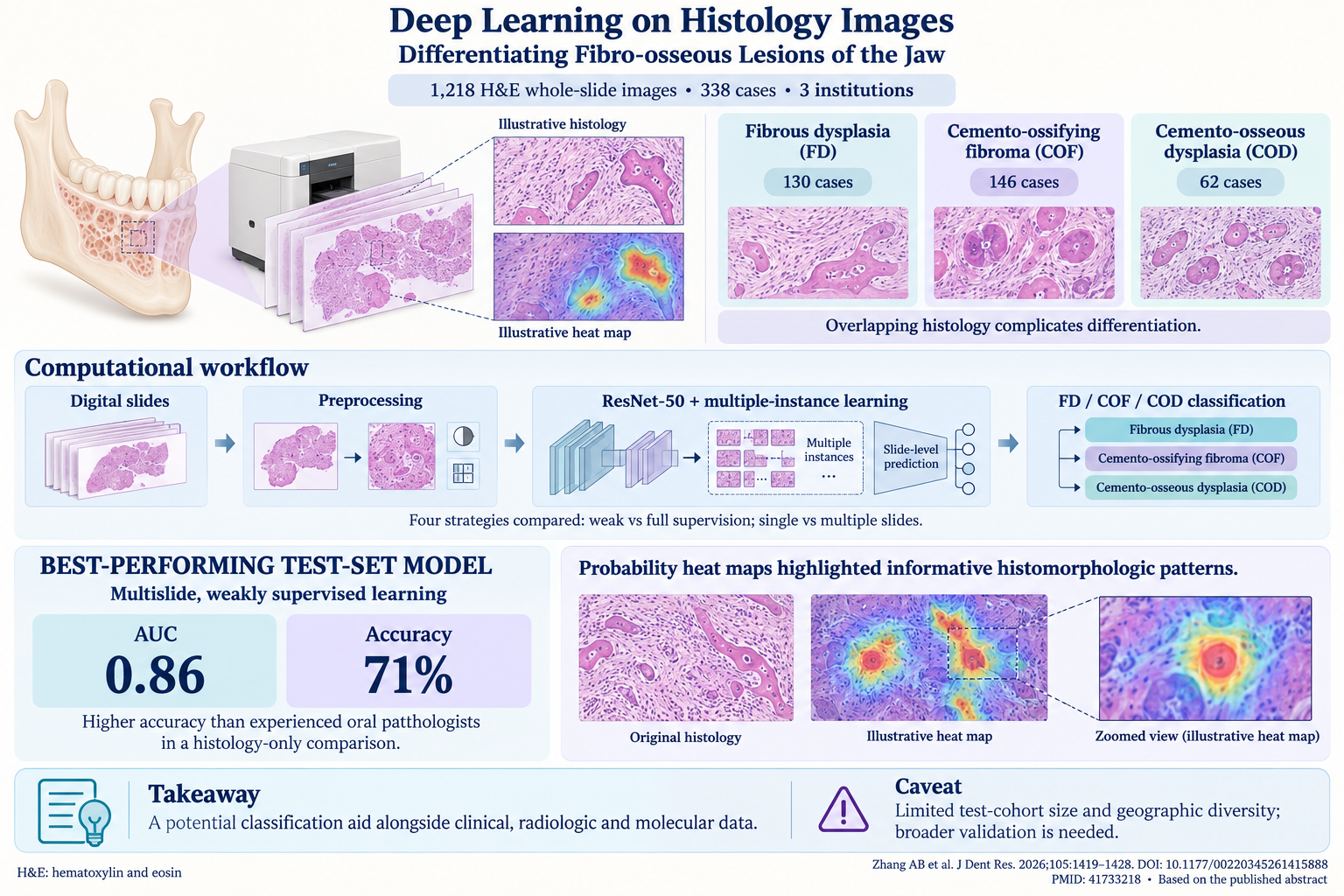
Fibrous dysplasia (FD), cemento-ossifying fibroma (COF), and cemento-osseous dysplasia (COD) are histologically similar jaw lesions that pose diagnostic challenges in pathology practice. A deep learning model using ResNet-50 and weakly supervised multislide training demonstrated superior performance over experienced oral pathologists, achieving an AUC of 0.86 and 71% accuracy. The model’s visual probability heatmaps pinpoint key distinguishing histomorphologic features, highlighting its potential as a clinical adjunct to traditional diagnostic methods.
Background
Fibro-osseous lesions of the jaw, including FD, COF, and COD, represent a group of benign but clinically diverse conditions characterized by the replacement of normal bone with fibrous tissue and varying degrees of mineralized material. These lesions exhibit overlapping histopathologic features, which frequently confound routine diagnostic efforts by oral pathologists. Accurate differentiation is critical as each entity has distinct clinical behavior, prognosis, and therapeutic implications. FD, typically a developmental abnormality, may stabilize or regress, while COF often requires surgical excision due to growth potential, and COD necessitates conservative management owing to its benign and asymptomatic nature.
Conventional diagnosis relies on morphology assessment by oral pathologists supplemented by clinical and radiographic data. However, given their histologic similarities, interobserver variability remains a concern, possibly leading to suboptimal clinical decisions. In recent years, artificial intelligence (AI), particularly deep learning applied to digital pathology, has emerged as a promising strategy to enhance diagnostic accuracy and consistency by objectively analyzing complex image patterns at scale.
Study Design
This retrospective, multicenter study aggregated a cohort of 338 cases classified as FD (n=130), COF (n=146), and COD (n=62) from three institutions. A total of 1,218 hematoxylin and eosin (H&E) stained whole slide images (WSI) were digitized and subjected to image preprocessing steps for standardization.
The study employed a ResNet-50 convolutional neural network architecture as the backbone for deep learning classification. Four different training strategies were evaluated: (1) fully supervised, single-slide; (2) fully supervised, multislide; (3) weakly supervised, single-slide; and (4) weakly supervised, multislide, with particular emphasis on multiple-instance learning frameworks that aggregate slide-level predictions to case-level diagnosis.
The primary endpoint was diagnostic performance measured by the area under the receiver operating characteristic curve (AUC) and accuracy in an independent test set. The model’s results were benchmarked against expert oral pathologists’ diagnostic assessments based solely on histology slides without clinical context.
Key Findings
Among the four strategies, the weakly supervised multislide model achieved the highest classification performance with an AUC of 0.86 and accuracy of 0.71 in the test set. This performance exceeded that of experienced oral pathologists, underscoring the model’s superior discriminative capability when interpreting histologic images alone.
Visualization using probability heatmaps demonstrated that the model successfully localized and emphasized histomorphologic regions critical for lesion differentiation. These regions included variations in fibrous stroma cellularity, the pattern and density of ossification, and the relationship of mineralized components to fibrous tissue, which are known diagnostic hallmarks in conventional pathology practice.
Importantly, the model’s performance reflected the incremental value of integrating multiple slides per case, simulating real-world diagnostic scenarios where pathologists review several sections to reach consensus.
Despite these encouraging results, the study acknowledges limitations, including the relatively modest sample size and restricted geographic representation, which might limit the model’s generalizability. Diverse patient populations and scanning platforms should be included in future research to validate robustness and clinical utility.
Expert Commentary
This investigation represents a significant advance in applying AI to oral pathology, demonstrating that deep learning models can not only match but potentially surpass experienced pathologists in challenging diagnostic tasks. The use of a weakly supervised approach with multiple-instance learning efficiently leverages slide-level labels without necessitating exhaustive pixel-level annotations, which are resource-intensive.
However, AI tools should be viewed as complementary aids rather than replacements for human expertise. Clinical, radiologic, and molecular data remain indispensable for comprehensive lesion evaluation. Future studies might explore multimodal AI models integrating histology with radiographic imaging and genetic markers to further refine diagnostic accuracy and personalized management.
Conclusion
This study confirms the feasibility and clinical promise of a weakly supervised multislide deep learning model in differentiating fibro-osseous lesions of the jaw using histologic images. By surpassing expert pathologists in accuracy using histology alone, this approach can serve as a robust adjunct to traditional pathology workflows, potentially reducing diagnostic ambiguity and improving patient management. Expanding the dataset with geographically diverse cases and incorporating multimodal diagnostic data will be critical next steps to establish widespread applicability and real-world impact.
Reference
Zhang AB, Li PY, Xue J, Zhang JY, You Z, Ge SH, Xu ZX, Sun ZP, Chang DX, Sun LS, Li TJ. Deep Learning on Histology Images for Differentiating Fibro-osseous Lesions of the Jaw. J Dent Res. 2026 Oct;105(11):1419-1428. doi: 10.1177/00220345261415888. Epub 2026 Feb 24. PMID: 41733218.