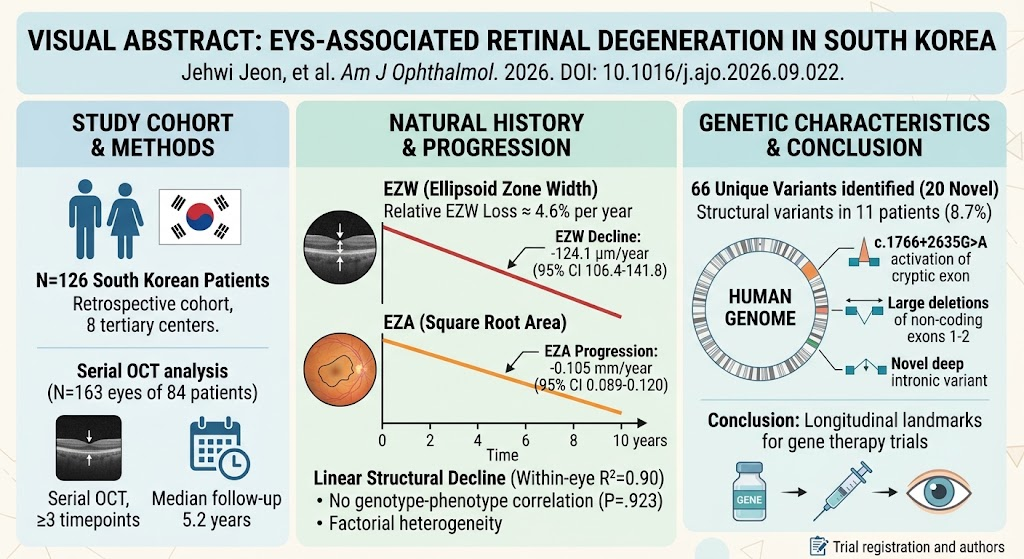
Highlight
This multicenter retrospective cohort study of 126 South Korean patients delineates the genetic and phenotypic spectrum of EYS-associated retinal degeneration (EYS-RD), quantifies longitudinal structural and functional progression, and reveals significant phenotypic heterogeneity without a clear genotype-phenotype correlation.
Key findings include a linear annual decline in ellipsoid zone width (EZW) of 124.1 µm, square root-transformed ellipsoid zone area (EZA) reduction by 0.105 mm, and best-corrected visual acuity (BCVA) worsening by 0.043 logMAR. The study identified 66 unique EYS variants, including 20 novel mutations, and highlights deep intronic and structural variants contributing to disease.
Study Background
Inherited retinal degenerations (IRDs) are a heterogeneous group of progressive diseases characterized by photoreceptor loss leading to visual impairment and blindness. Among these, EYS (eyes shut homolog) gene mutations represent a major cause of autosomal recessive retinitis pigmentosa particularly in Asian populations. Despite its significance, comprehensive natural history data detailing the genetic spectrum, clinical phenotype, and disease progression kinetics remain limited in East Asian cohorts.
Given the emerging landscape of gene and molecular therapies targeting inherited retinal disorders, establishing quantitative, longitudinal benchmarks of retinal structural and functional decline is critical for optimizing clinical trial endpoints and therapeutic timing. Furthermore, understanding the phenotypic variability and genotype-phenotype relationships can aid patient prognostication and precision medicine approaches.
Study Design
This retrospective multicenter cohort study was conducted across eight tertiary referral centers in South Korea, enrolling 126 patients with biallelic EYS mutations confirmed by genetic testing. The cohort included equal proportions of females and males (50% each) with a median follow-up time of 5.2 years (interquartile range 1.2–9.0 years).
Longitudinal clinical data encompassed best-corrected visual acuity (BCVA) measured in logMAR units and spectral-domain optical coherence tomography (SD-OCT) imaging focusing on ellipsoid zone width (EZW) and ellipsoid zone area (EZA). A total of 163 eyes from 84 patients were eligible for serial OCT analysis, with most eyes having three or more imaging time points, yielding 591 observations of EZW measurements.
Progression rates for EZW, EZA, and BCVA were modeled using three-level linear mixed-effects regression to account for repeated measures nested within eyes and patients. Genotype stratification compared biallelic loss-of-function (LoF) variants versus genotypes harboring at least one missense allele.
Key Findings
The study identified 66 unique EYS variants in this cohort, including 20 novel mutations. Structural variants, such as large deletions affecting non-coding exons 1–2 and a deep intronic variant (c.1766+2635G>A) activating a cryptic exon, were found in approximately 8.7% of patients. This comprehensive genotyping underscores the complexity of EYS mutational mechanisms in retinal degeneration.
Longitudinal analysis revealed a consistent linear decline in EZW with an annual reduction of 124.1 µm (95% confidence interval [CI], 106.4–141.8 µm/year). Similarly, the square root-transformed EZA decreased by 0.105 mm/year (95% CI, 0.089–0.120 mm/year). BCVA worsened at a rate of 0.043 logMAR per year (95% CI, 0.025–0.060 logMAR/year), corresponding to a gradual loss of visual function.
The linear model demonstrated excellent goodness-of-fit, with a median within-eye R² of 0.900 and no significant improvement when incorporating quadratic time terms (P=0.239), indicating steady progressive retinal degeneration over the observation period.
Importantly, no statistically significant difference in progression was observed between patients with biallelic LoF genotypes and those carrying at least one missense variant (time × genotype interaction, P=0.923), suggesting that genotype alone does not predict disease trajectory.
Expert Commentary
This study significantly advances the understanding of EYS-associated retinal degeneration by providing robust, quantitative natural history data and expanding the known genetic spectrum in a large East Asian cohort. The demonstration of a linear retinal structural decline simplifies modeling disease progression and supports the use of ellipsoid zone measurements as sensitive biomarkers for clinical trials. These benchmarks can help calibrate expectations for disease course and therapeutic response assessment.
The lack of genotype-phenotype correlation highlights the considerable phenotypic heterogeneity in EYS-RD and implies that additional genetic modifiers, environmental factors, or epigenetic mechanisms may influence disease severity and progression. This complexity underlines the necessity for individualized patient evaluation and cautious interpretation of genetic findings.
Despite its strengths, the retrospective design and heterogeneous follow-up intervals warrant careful consideration when extrapolating progression rates. Furthermore, functional measures beyond BCVA, such as microperimetry or electrophysiology, could enrich assessment of visual function decline.
Conclusion
This large-scale, multicenter cohort study establishes essential quantitative reference points for retinal structural and visual function deterioration in South Korean patients with EYS-associated retinal degeneration. The delineation of 66 causative variants, including novel and structural types, broadens the mutational landscape and informs genetic diagnostics.
Linear progression rates of EZW and EZA of approximately 4.6% annual loss reinforce their utility as reliable endpoints for future gene therapy and interventional trials targeting EYS-RD. The absence of a clear genotype-phenotype correlation suggests multifactorial influences on disease phenotype and progression, advocating for further research into modifying factors.
Collectively, these findings enhance disease understanding, support patient counseling, and pave the way for optimized clinical trial design in EYS-related inherited retinal degeneration.
Funding and Clinical Trials
The study was conducted across national tertiary care centers in South Korea and supported by institutional funding. There was no mention of clinical trial registration numbers or specific external funding bodies in the source publication.
References
Jeon J, Surl D, Park H, Lee J, Kim YJ, Sangermano R, Won D, Lee ST, Kim M, Byeon SH, Kim SS, Yang JM, Ahn SJ, Kim JH, Ahn J, Sagong M, Lee JY, Joo G, Woo SJ, Bujakowska K, Han J, Lee CS. EYS-associated retinal degeneration in South Korea: Genetic, Phenotype, and Natural History. Am J Ophthalmol. 2026 Sep 23. PMID: 42777796.
Additional references for contextual background and methodology are available upon request.