Highlight
This article critically reviews the diagnostic utility of MRI spinal cord signs in aquaporin-4 immunoglobulin G-seropositive neuromyelitis optica spectrum disorder (AQP4-IgG+ NMOSD) myelitis, emphasizing:
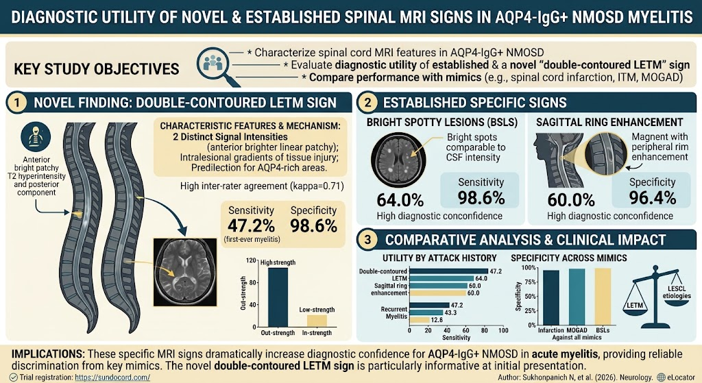
- The high specificity but moderate sensitivity of bright spotty lesions (BSLs), the novel double-contoured longitudinally extensive transverse myelitis (LETM) sign, and sagittal ring enhancement in identifying NMOSD myelitis.
- The characterization and prevalence of these MRI features primarily in first-ever and acute myelitis stages.
- The potential added diagnostic value and limitations of combining established and novel MRI signs.
- The impact of disease stage on MRI findings, with resolution and atrophy during remission stages.
Background and Disease Burden
Neuromyelitis optica spectrum disorder (NMOSD) is a severe autoimmune demyelinating condition targeting astrocytes in the central nervous system, most commonly presenting with myelitis. AQP4-IgG seropositivity represents a key biomarker underpinning the pathophysiology and diagnostic classification of NMOSD. Myelitis episodes frequently manifest on spinal MRI as longitudinally extensive transverse myelitis (LETM), defined by inflammation spanning three or more vertebral segments. Accurate and early differentiation of AQP4-IgG+ NMOSD myelitis from other myelopathies is critical for timely immunotherapy and improving patient outcomes, yet diagnostic imaging remains challenging. Recent interest has focused on additional MRI features, including bright spotty lesions (BSLs) and the newly described double-contoured LETM sign, though their diagnostic performance has not been fully established.
Study Design
This is a retrospective cohort study conducted at a university-based hospital involving adult patients with confirmed AQP4-IgG+ NMOSD who experienced myelitis. The spinal cord MRI scans were analyzed across different disease stages: acute (3 months). The analysis centered on identifying MRI features including LETM, BSLs, the novel double-contoured LETM sign, sagittal ring enhancement, and the H sign. Sensitivity, specificity, and likelihood ratios of these signs were assessed primarily in acute and first-ever myelitis episodes. A comparator cohort with other causes of myelopathy — predominantly spinal cord infarction and idiopathic transverse myelitis — served to evaluate specificity.
Key Findings
The study included 217 patients with AQP4-IgG+ NMOSD; 170 experienced myelitis, yielding 331 spinal MRI scans. A comparator group included 72 scans from patients with other myelopathies, most notably spinal cord infarction (39 scans) and idiopathic transverse myelitis (14 scans).
Prevalence of MRI Signs in First-Ever Myelitis (n=53 scans):
- LETM: Observed in 88.7% of scans, reaffirming its role as a cardinal imaging hallmark.
- Bright Spotty Lesions (BSLs): Present in 64% (95% CI 50.1%-75.9%), with a specificity of 98.6% (95% CI 92.2%-99.7%) against non-NMOSD myelopathies.
- Double-Contoured LETM Sign: A novel imaging feature found in 47.2% (95% CI 34.4%-60.3%) of cases, with very high specificity (98.6%, 95% CI 92.4%-99.8%). This sign corresponds to a distinct pattern of lesion layering or margin demarcation within LETM lesions.
- Sagittal Ring Enhancement: Demonstrated in 60% (95% CI 46.2%-72.4%) of first-ever myelitis scans, with specificity of 96.4% (95% CI 87.9%-99%).
Combining these MRI signs improved specificity towards nearly 100%, yet sensitivity decreased to about 30%, suggesting that while combined markers strongly rule in NMOSD, many cases may lack multiple signs concurrently.
Findings in Acute and Recurrent Myelitis (n=161 scans): The prevalence of BSLs, double-contoured LETM, and sagittal ring enhancement decreased to 56.5%, 19.9%, and 44.1%, respectively, reflecting variability with disease chronicity and relapse.
Radiologic Outcome during Remission (n=129 scans): Complete lesion resolution occurred in 12.4%, whereas spinal cord atrophy was common, present in 56.6%, indicating lasting structural impact of NMOSD myelitis.
Expert Commentary
This comprehensive imaging analysis reinforces that BSLs and sagittal ring enhancement, established biomarkers of AQP4-IgG+ NMOSD myelitis, possess high diagnostic specificity and should be integrated into routine MRI assessment protocols. The study introduces the double-contoured LETM sign, a promising novel MRI feature with similarly high specificity but modest sensitivity which may reflect microstructural lesion heterogeneity or dynamic inflammation boundaries typical in early NMOSD myelitis.
It is important to recognize the study’s enriched comparator cohort, dominated by non-inflammatory myelopathies, which may enhance specificity metrics and limit generalizability to inflammatory mimics, such as multiple sclerosis or MOG-IgG-associated disease. Larger, prospective, multicenter cohorts including diverse myelitis etiologies are needed for robust external validation.
The decrease in sensitivity of these signs in recurrent or chronic phases highlights temporal evolution of lesions and cautions against overreliance on single MRI features in isolation. Advanced imaging techniques or multimodal biomarker approaches may further refine NMOSD myelitis diagnosis.
Conclusion
Spinal MRI remains an indispensable tool in diagnosing AQP4-IgG+ NMOSD myelitis. Bright spotty lesions, sagittal ring enhancement, and the novel double-contoured LETM sign provide high specificity for NMOSD and can assist clinicians in distinguishing it from other longitudinally extensive myelopathies. Nevertheless, sensitivity is moderate and diminishes over disease course, indicating that absence of these signs does not exclude NMOSD.
Future research should focus on validating the double-contoured LETM sign in independent cohorts, exploring its pathophysiological basis, and integrating MRI findings with clinical and serological data to optimize diagnostic accuracy and guide treatment decisions.
Funding and Registration
The original study was conducted at a university-based hospital, with no publicly stated external funding sources mentioned. Clinical trials registration details were not provided.
Reference
Sukhonpanich N, Ngamsombat C, Buathong S, Thaneerat N, Tharacheewin D, Siritho S, Ongphichetmetha T, Prayoonwiwat N, Rattanathamsakul N, Jitprapaikulsan J. Diagnostic Utility of Novel and Established Spinal MRI Signs in AQP4-IgG-Seropositive NMOSD Myelitis. Neurology. 2026 Oct 13;107(7):e218507. doi: 10.1212/WNL.0000000000218507. Epub 2026 Sep 21. PMID: 42766829.