
Highlight
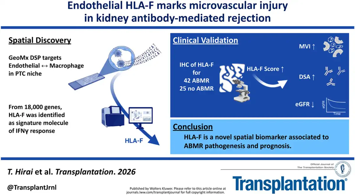
1. Spatial transcriptomic profiling of kidney allograft biopsies reveals an interferon-γ-driven molecular signature in the peritubular capillary (PTC) microenvironment during antibody-mediated rejection (ABMR).
2. HLA-F, expressed in CD31+ endothelial cells within the PTC niche, emerges as a distinct and prognostically significant marker for ABMR.
3. High endothelial HLA-F expression correlates with microvascular inflammation severity, donor-specific antibody (DSA) positivity, and subsequent decline in allograft kidney function.
4. Spatially resolved detection of HLA-F offers potential to enhance diagnostic precision and risk stratification in kidney transplant rejection.
Study Background
Kidney transplantation remains the optimal therapeutic option for patients with end-stage renal disease, yet allograft rejection, particularly antibody-mediated rejection (ABMR), limits graft survival. ABMR is characterized histopathologically by microvascular inflammation (MVI), including capillaritis and glomerulitis, caused by antibodies targeting donor human leukocyte antigen (HLA) molecules. However, MVI alone, despite being essential for diagnosis, lacks sufficient specificity and prognostic accuracy, posing challenges to timely individualized management. There is a clinical need for molecular biomarkers that directly reflect the endothelial and immune cell interactions underpinning ABMR pathology to improve diagnosis and prognostication. Recent advances in spatial transcriptomic technologies enable precise molecular characterization of spatially defined microenvironments like peritubular capillaries (PTCs), critical sites of rejection injury. Against this background, the study by Hirai et al. explores endothelial gene expression signatures within the PTC niche, aiming to identify novel biomarkers linked to ABMR pathogenesis and outcome.
Study Design
The investigation comprised two principal phases. In the discovery phase, the authors analyzed kidney allograft biopsy samples using spatial transcriptomics via the GeoMx Digital Spatial Profiler. Targeting the PTC niche, they performed microdissection guided by immunostaining for CD31 (endothelial cells) and CD68 (macrophages) in six biopsies (3 with ABMR and 3 with T cell-mediated rejection, TCMR). RNA was extracted from these spatially defined regions and subjected to whole-transcriptome sequencing (Whole Transcriptome Atlas, WTA), interrogating over 18,000 genes. Gene set enrichment analysis focused on interferon-related pathways and candidate molecules differentiating ABMR versus TCMR.
In the validation phase, immunohistochemical (IHC) analyses for HLA-F were conducted on a larger cohort of 67 kidney transplant biopsies (42 ABMR and 25 non-ABMR control samples). Correlations between endothelial HLA-F expression, MVI scores, donor-specific antibody (DSA) status, and clinical outcomes including kidney function trajectories post-biopsy were assessed.
Key Findings
Spatial transcriptomic profiling demonstrated that both CD31+ endothelial cells and CD68+ macrophages within the PTC microenvironment shared a coordinated interferon-γ-driven transcriptional program in ABMR. This transcriptional signature, not observed in TCMR samples, included marked upregulation of the non-classical HLA class I gene HLA-F in endothelial cells.
HLA-F expression in CD31+ endothelial cells was validated by IHC in the larger cohort. Key correlations included:
- Microvascular inflammation severity: HLA-F protein levels positively correlated with extent of peritubular capillaritis and glomerulitis, hallmarks of MVI.
- Donor-specific antibody positivity: Elevated endothelial HLA-F was associated with presence of circulating DSA, supporting its link to alloimmune activation.
- Prognosis: Among patients with active ABMR, those with higher HLA-F expression exhibited significantly greater decline in estimated glomerular filtration rate (eGFR) after biopsy, indicating worse allograft function over time.
Collectively, these findings establish HLA-F as a novel, spatially localized molecule in the PTC niche that reflects endothelial immune activation driven by interferon signaling in ABMR and predicts adverse graft outcomes.
Expert Commentary
The study by Hirai et al. leverages cutting-edge spatial transcriptomic technology to overcome limitations of conventional histopathology, providing molecular granularity about the endothelial-immune interface in kidney ABMR. Identification of HLA-F as a prognostic biomarker is particularly intriguing given its role as a non-classical HLA molecule implicated in immune regulation and tolerance. The detection of an interferon-γ-driven endothelial program further substantiates the centrality of type II interferons in ABMR pathogenesis.
Despite compelling data, some limitations warrant consideration. The initial discovery was based on a small sample size, which might limit the breadth of gene expression variability captured. Validation in larger, multi-center cohorts and exploration of HLA-F’s mechanistic role—whether it actively modulates immune interactions or primarily serves as a marker—are needed. Additionally, the potential for HLA-F to guide therapeutic decisions or monitor treatment response remains to be explored.
From a clinical perspective, spatially resolved biomarkers such as endothelial HLA-F could refine diagnostic accuracy beyond morphology and DSA testing alone, enabling personalized risk stratification and tailored immunosuppressive strategies. Integration with non-invasive approaches, including circulating biomarkers, may further optimize transplant monitoring.
Conclusion
This study identifies endothelial HLA-F expression in the peritubular capillary niche as a novel and promising biomarker for ABMR in kidney transplantation. HLA-F is closely linked to microvascular injury severity, alloantibody presence, and functional deterioration post-rejection diagnosis. Spatial transcriptomics provides a powerful platform to unravel the molecular microenvironment underlying graft injury, offering potential utility for improved diagnostics and personalized prognosis assessment in kidney allograft rejection. Future studies should validate these findings in larger cohorts and assess the clinical utility of HLA-F-guided management strategies to improve long-term transplant outcomes.
Funding and ClinicalTrials.gov
The original study did not specify funding sources within the abstract. No clinical trial registration was detailed.
References
1. Hirai T, Shimizu T, Fukuda H, Unagami K, Omoto K, Inui M, Ishida H, Takagi T. Spatial Analysis Identifies Endothelial HLA-F as a Prognostic Marker in Antibody-mediated Rejection. Transplantation. 2026 Jun 17;110(11):e2284-e2299. PMID: 42302751.
2. Haas M. The Banff 2017 Kidney Meeting Report: Revised diagnostic criteria for chronic active T cell–mediated rejection, antibody-mediated rejection, and prospects for integrative diagnostics. Am J Transplant. 2018 Jan;18(1):293-307.
3. Colvin RB. Antibody-mediated renal allograft rejection: diagnosis and pathogenesis. J Am Soc Nephrol. 2007 Dec;18(4):1046-56.
4. Wiebe C, Nickerson PW. Understanding antibody-mediated rejection: progress and opportunities. Curr Opin Organ Transplant. 2017 Dec;22(6):425-432.
Specialty calculators
For clinical reference; use professional judgment.