Highlight
– Hemorrhagic vocal fold polyps demonstrate significantly higher complete regression rates following office-based blue laser therapy combined with intralesional steroid injection compared to nonhemorrhagic polyps.
– Both hemorrhagic and nonhemorrhagic polyps showed improvement in Voice Handicap Index-10 (VHI-10) scores post-treatment, with a trend toward greater vocal benefit in hemorrhagic polyps.
– Jitter, an acoustic parameter reflecting voice stability, was significantly better improved in patients with hemorrhagic polyps.
– The combined minimally invasive approach offers an effective outpatient treatment option with a mean follow-up period of approximately 13 weeks.
Study Background
Vocal fold polyps are common benign lesions that affect phonation and voice quality. They can be broadly classified into hemorrhagic and nonhemorrhagic types based on their morphological and vascular characteristics. Hemorrhagic polyps typically contain extravasated blood, often resulting from phonotrauma, and exhibit a distinct red or bluish hue, whereas nonhemorrhagic polyps are generally fibrotic or edematous.
Historically, treatment options for vocal fold polyps range from conservative voice therapy to surgical excision under general anesthesia. However, the advent of office-based laser procedures combined with steroid injections offers a less invasive alternative with reduced recovery time. Blue laser, with its favorable tissue targeting and hemostatic properties, is increasingly utilized for such interventions. Yet, there is limited evidence evaluating whether polyp morphology influences treatment outcomes with this modality.
Study Design
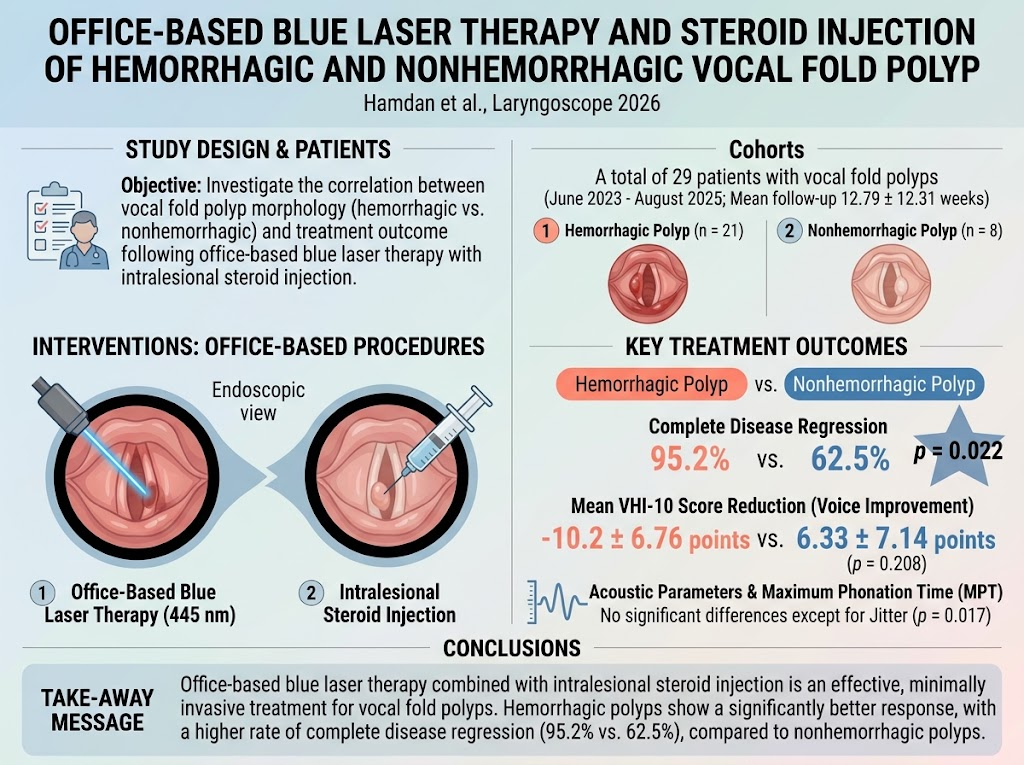
This retrospective cohort study analyzed 29 patients diagnosed with vocal fold polyps treated at a tertiary referral center between June 2023 and August 2025. The intervention consisted of office-based blue laser therapy combined with intralesional steroid injection delivered under endoscopic guidance.
Patients were classified into two subgroups based on polyp morphology observed on video recordings: hemorrhagic (n=21) and nonhemorrhagic (n=8). Baseline demographic characteristics were comparable between groups.
Primary endpoints included subjective voice outcomes measured by the Voice Handicap Index-10 (VHI-10), acoustic analysis parameters including jitter, shimmer, and fundamental frequency, maximum phonation time (MPT), and objective observation of disease regression assessed through endoscopic follow-up. The mean follow-up duration was 12.79 ± 12.31 weeks.
Key Findings
Both subgroups exhibited improvement after treatment, with mean VHI-10 scores decreasing significantly: hemorrhagic polyps improved by 10.2 ± 6.76 points, and nonhemorrhagic polyps by 6.33 ± 7.14 points. Although the difference in subjective voice improvement was not statistically significant (p = 0.208), it suggested a trend favoring hemorrhagic polyps.
Critically, the rate of complete disease regression was significantly higher in the hemorrhagic polyp group (95.2%) compared with the nonhemorrhagic group (62.5%) (p = 0.022). This finding indicates stronger treatment responsiveness of hemorrhagic polyps to blue laser therapy with steroid injection.
Acoustic parameters largely showed no significant intergroup differences, except for jitter, which improved significantly more in the hemorrhagic group (p = 0.017). Jitter reflects frequency stability of voice and its improvement suggests enhanced vocal cord vibration regularity post-treatment.
MPT and shimmer did not differ significantly between groups, indicating comparable improvements in breath control and amplitude perturbation regardless of polyp morphology.
No major adverse events or complications were reported, underscoring the safety and tolerability of this minimally invasive approach.
Expert Commentary
The study provides important clinical insights supporting the differential response of vocal fold polyps based on morphological subtype to office-based blue laser treatment combined with steroid injection. Hemorrhagic polyps, by virtue of their vascular content, may be more amenable to coagulative effects of the blue laser, promoting more effective lesion resolution.
Intralesional steroid administration likely augments outcomes by reducing local inflammation and fibrosis, facilitating restoration of normal vocal fold function.
Limitations include the relatively small sample size, particularly for the nonhemorrhagic subgroup, and the retrospective design. Additionally, the mean follow-up of approximately 13 weeks provides short- to mid-term outcomes; long-term efficacy and recurrence rates remain to be evaluated.
Inclusion of voice therapy adjuncts and standardized voice outcome measures could improve assessment robustness in future prospective studies.
Comparison with traditional surgical excision outcomes would also clarify the relative benefits of office-based laser approaches.
Conclusion
Office-based blue laser therapy combined with intralesional steroid injection represents a promising, minimally invasive treatment for vocal fold polyps. This study demonstrates that hemorrhagic polyps respond significantly better in terms of complete regression and voice quality improvement than nonhemorrhagic polyps.
Clinicians should consider polyp morphology when selecting candidates for this intervention and counseling patients on expected outcomes. Further large-scale, prospective research with longer follow-up is warranted to confirm these findings and optimize treatment protocols.
Funding and Clinical Trials
The study was conducted at a tertiary referral center with no external funding reported. Clinical trial registration information was not provided.
References
1. Hamdan AL, Abou Raji Feghali P, Ghzayel L, et al. Office-Based Blue Laser Therapy and Steroid Injection of Hemorrhagic and Nonhemorrhagic Vocal Fold Polyp. Laryngoscope. 2026;136(10):4401-4407. doi:10.1002/lary.30850
2. Cohen MT, Slavit DH. Office-based treatment of vocal fold lesions: Current perspectives. Curr Opin Otolaryngol Head Neck Surg. 2019;27(6):425-430. doi:10.1097/MOO.0000000000000583
3. Mor A, Misono S. Diagnosing and managing vocal fold hemorrhage and polyps. Otolaryngol Clin North Am. 2020;53(3):395-405. doi:10.1016/j.otc.2020.02.001
4. Woo P, Casper J. Advances in office-based procedures for benign vocal fold lesions. Otolaryngol Clin North Am. 2018;51(6):1093-1105. doi:10.1016/j.otc.2018.07.005