Early Optic Nerve Head Swelling Rate Predicts Visual Outcome Deterioration in Nonarteritic Anterior Ischemic Optic Neuropathy: An Evidence-Based Review
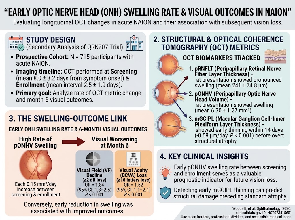
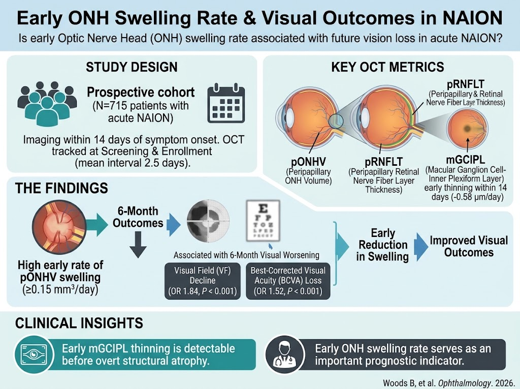
Rapid early optic nerve head swelling, quantified by peripapillary optic nerve head volume increase, strongly predicts visual field and acuity worsening in acute NAION.
Macular ganglion cell-inner plexiform layer thinning is detectable within two weeks of symptom onset, preceding overt structural optic nerve atrophy.
Optical coherence tomography angiography demonstrates a sequential decline in peripapillary and macular vascular densities paralleling structural damage progression in NAION.
Combined OCT and scanning laser polarimetry modalities reveal distinct yet complementary insights on axonal integrity during acute NAION-induced optic nerve head swelling.