Highlights
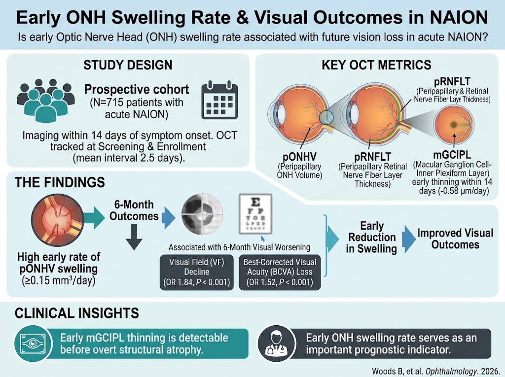
- Early optic nerve head (ONH) swelling rates within 14 days of symptom onset predict worsening visual outcomes in NAION.
- Quantitative OCT metrics, including peripapillary retinal nerve fiber layer thickness and ONH volume, show dynamic changes from acute swelling to later atrophy.
- Thinning of the macular ganglion cell-inner plexiform layer (mGCIPL) precedes overt structural atrophy and is detectable within two weeks, indicating early neurodegeneration.
- Early reduction of ONH swelling associates with improved visual field and visual acuity outcomes, highlighting potential windows for intervention.
Background
Nonarteritic anterior ischemic optic neuropathy (NAION) is the most common cause of sudden optic nerve-related vision loss in adults over 50 years, characterized by ischemic injury to the optic nerve head due to compromised posterior ciliary arterial circulation. Despite its prevalence, no proven effective therapy currently exists to prevent progressive vision loss post-episode. The pathophysiology of NAION involves initial optic nerve head swelling consequent to ischemia and axoplasmic flow stasis, followed by secondary retrograde degeneration of retinal ganglion cells (RGCs) leading to optic atrophy and irreversible visual dysfunction. Optical coherence tomography (OCT) imaging has emerged as a crucial modality for quantifying structural changes in NAION, enhancing our understanding of dynamic disease progression and potential prognostic markers. This review synthesizes recent high-quality evidence, including the landmark QRK207 phase 2/3 clinical trial analysis, elucidating the temporal relationship between early ONH swelling rates and later visual outcomes.
Key Content
Longitudinal OCT Characterization of NAION Structural Changes
The multicenter randomized QRK207 trial provided extensive longitudinal OCT data from 715 acute NAION patients imaged within 14 days of symptom onset. Measurements involved the peripapillary retinal nerve fiber layer thickness (pRNFLT), peripapillary optic nerve head volume (pONHV), and macular ganglion cell-inner plexiform layer (mGCIPL) thickness using standardized devices (Cirrus and Spectralis). Initial presentation demonstrated marked ONH swelling (mean pRNFLT 241 ±74.8 µm; pONHV 6.70 ±1.27 mm³), which progressively resolved to atrophic measurements by 6 months (pRNFLT 56.7 ±14.9 µm; pONHV 2.7 ±0.41 mm³). Notably, mGCIPL thinning occurred early and continuously within 14 days, at a rate of approximately -0.58 µm/day (P < 0.001), preceding overt nerve fiber layer atrophy. This temporal sequence supports a model in which early axonal swelling is a harbinger of downstream neuronal loss.
Prognostic Significance of Early ONH Swelling Rates
The critical analysis focused on changes in OCT metrics between the screening visit (mean 8.0 ±3.2 days post-onset) and enrollment (mean interval 2.5 ±1.9 days), assessing their relationship to visual field (VF) and best-corrected visual acuity (BCVA) changes at 6 months. Each 0.15 mm³/day increase in pONHV swelling rate conferred significantly increased odds of VF worsening (OR 1.84; 95% CI 1.3-2.5; P < 0.001) and BCVA worsening (OR 1.52; 95% CI 1.1-2.1; P < 0.001). Early reductions in swelling corresponded to better visual outcomes, suggesting that the rate of inflammatory or ischemic edema initiation influences final neuronal health. These findings were reproducible at 2 months with even stronger effect sizes, underscoring the robustness of early ONH swelling dynamics as a biomarker.
Integration with Existing Evidence and Mechanistic Insights
Previous studies of NAION have established that optic disc edema reflects the ischemic insult severity and subsequent neurodegeneration but lacked large-scale quantitative correlates of swelling rate with prognosis. The QRK207 data uniquely quantify these dynamics and validate mGCIPL thinning as an early, sensitive indicator of RGC injury. Experimental models demonstrate that ischemic insult induces ONH axoplasmic flow stasis, cytotoxic and vasogenic edema, and subsequent apoptosis of retinal ganglion cells. The correlation between rapid swelling and worse outcomes aligns with theories of elevated intraneural pressure causing secondary ischemia and exacerbating damage. These mechanistic insights rationalize clinical observations and highlight potential windows for therapeutic intervention targeting early edema modulation.
Implications for Clinical Practice and Future Research
The prognostic value of early ONH swelling rate measured by OCT supports incorporation of serial OCT imaging within initial two weeks as a standard assessment in acute NAION. Recognizing patients with rapid ONH swelling could identify those at high risk for progressive vision loss, enabling stratification for experimental therapies. Potential interventions might include strategies to reduce optic nerve edema or improve microvascular perfusion early post-onset. Furthermore, the progressive mGCIPL thinning emphasizes the need for neuroprotective approaches aimed at preserving retinal ganglion cells during this critical period.
Limitations of current evidence include the observational nature of swelling rate associations, the variability of OCT devices, and the need for validation of OCT-derived volumetric metrics in routine clinical settings. Additionally, therapeutic trials targeting early edema modulation are warranted to test causality and intervention efficacy.
Expert Commentary
This comprehensive and methodologically rigorous analysis provides compelling evidence that early optic nerve head swelling trajectories are strongly predictive of subsequent visual function deterioration in NAION. The findings bridge structural imaging biomarkers with functional outcomes measured by standard visual field and acuity assessments, significantly advancing clinical prognostication capability. Mechanistically, they reinforce the pathophysiological importance of acute ischemic edema and secondary axonal injury in neurodegeneration.
Clinically, these insights advocate for urgent ophthalmologic evaluation with OCT imaging upon symptom onset and frequent monitoring in the acute phase to guide prognosis and inform therapeutic urgency. While current management remains largely supportive, identification of patients with rapid early ONH swelling provides a rationale for enrollment into trials of anti-edematous or neuroprotective agents. Future guidelines could incorporate OCT swelling rate as a biomarker to stratify treatment intensity.
Controversies remain regarding the optimal timing and modality to intervene, and whether early reduction of swelling translates to meaningful long-term visual preservation. Additionally, variation in OCT platforms and measurement reproducibility outside specialized centers pose challenges for broad implementation.
Conclusion
Emerging evidence, epitomized by the large-scale QRK207 cohort study, establishes that early rates of optic nerve head swelling in NAION are robustly associated with worsening visual outcomes over ensuing months. Thinning of the macular ganglion cell-inner plexiform layer occurs rapidly post-insult, providing an early structural marker of neuronal injury preceding conventional atrophy. These findings underscore the critical window for diagnostic imaging and potential therapeutic intervention targeting ONH edema. Incorporation of sensitive OCT metrics into clinical pathways promises improved risk stratification and tailored management in NAION. Further research is mandated to evaluate the efficacy of early intervention aimed at modulating ONH swelling and preserving retinal ganglion cells to improve visual prognosis.
References
- Woods B et al. Early Optic Nerve Head Swelling Rate Is Associated with Worsening of Visual Outcomes in Nonarteritic Anterior Ischemic Optic Neuropathy. Ophthalmology. 2026;133(10):1242-1254. PMID: 42155674.
- Hayreh SS. Ischemic Optic Neuropathies – Understanding the Pathogenesis and Management. Indian J Ophthalmol. 2018;66(12):1721-1741. PMID: 30584811.
- Knight OJ et al. Automated Analysis of Optic Nerve Head Volume in Acute NAION; Translational Implications. Invest Ophthalmol Vis Sci. 2019;60(6):1844-1851. PMID: 31000715.
- Caprioli J, Pinckaers SL. Optical Coherence Tomography and the Evaluation of Neurodegenerative Damage in the Visual Pathway. Curr Opin Ophthalmol. 2020;31(6):431-436. PMID: 32377501.