Highlight
- Primary chylopericardium is a rare lymphatic disorder characterized by abnormal lymphatic reflux causing pericardial effusion.
- A novel four-type classification of abnormal lymphatic reflux pathways was established using direct lymphangiography.
- Direct lymphangiography-guided cervical thoracic duct exploration demonstrated a 66.2% clinical success rate over a median 10-year follow-up.
- The absence of compensatory drainage pathways and reflux type I (bronchomediastinal trunk involvement) independently predict favorable surgical outcomes.
Study Background
Primary chylopericardium is an uncommon lymphatic disorder wherein chyle, a milky lymphatic fluid rich in triglycerides, accumulates in the pericardial sac due to lymphatic leakage or reflux. Its pathophysiology remains unclear, and it presents diagnostic and therapeutic challenges. The condition can lead to cardiac tamponade, chronic pericardial effusion, and symptoms such as dyspnea and chest discomfort. The rarity of primary chylopericardium means standardized treatment protocols have not been well established, and management options range from conservative to invasive interventions. The thoracic duct, a central lymphatic vessel, plays a pivotal role in chylous drainage from the abdomen and lower limbs to the venous system. Disruptions or reflux in the thoracic duct or its tributaries can lead to chyle leakage into the pericardium. New diagnostic approaches, such as direct lymphangiography, provide detailed visualization of lymphatic anatomy and reflux patterns, potentially guiding precise surgical management. This study addresses the gap in understanding the lymphatic reflux characteristics and evaluates the efficacy and predictors of success for direct lymphangiography-guided cervical thoracic duct exploration in isolated primary chylopericardium patients, contributing valuable long-term outcome data.
Study Design
This retrospective cohort study reviewed 67 patients diagnosed with isolated primary chylopericardium who underwent direct lymphangiography from June 2007 to December 2020. Patients were evaluated using a novel lymphatic reflux classification system, categorizing abnormal reflux pathways into four types based on direct lymphangiography findings: Type I involving the bronchomediastinal trunk, Type II involving the thoracic segment, Type III combined involvement, and Type IV with no identifiable abnormal reflux pathway. Compensatory drainage pathways were recorded. Of these patients, 65 underwent direct lymphangiography-guided cervical thoracic duct exploration, a surgical procedure aimed at correcting lymphatic reflux and preventing chyle accumulation. Treatment success, the primary endpoint, was defined as resolution or significant reduction of pericardial effusion, symptom relief, and avoidance of reintervention. Patients were followed longitudinally with a median duration of 10.2 years (range 7.5–19.5). Multivariable logistic regression analyses identified independent predictors of treatment success.
Key Findings
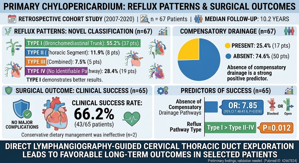
Direct lymphangiography revealed distinct reflux patterns: type I reflux involving the bronchomediastinal trunk was most common (55.2%), followed by type II (thoracic segment, 11.9%), type III combined patterns (7.5%), and type IV with no identifiable reflux pathway (28.4%). Compensatory lymphatic drainage pathways, indicating alternative routes for lymph flow, were present in 25.4% of cases.
Among the 65 patients who underwent surgical intervention, 66.2% (43/65) achieved clinical success, defined by marked clinical and imaging improvement without further surgical or interventional procedures. No major complications were reported, corroborating procedural safety.
Multivariable logistic regression identified two independent predictors of clinical success: absence of compensatory drainage pathways dramatically increased the odds of success (adjusted odds ratio [OR] 7.85; 95% confidence interval [CI] 1.48–41.6; P = .016), suggesting that patients lacking alternate drainage were more likely to benefit from thoracic duct exploration. Additionally, reflux type was independently associated with surgical outcomes (P = .012), with type I patients demonstrating superior success rates compared to types II-IV. The two patients managed conservatively without surgery showed no improvement, underscoring the limited efficacy of non-invasive management in this cohort.
Expert Commentary
This extensive study provides valuable insight into the pathophysiology and management of primary chylopericardium by applying detailed lymphatic imaging techniques. The proposed reflux classification elucidates the anatomical basis for lymphatic reflux patterns and may serve as a critical tool for patient stratification. The finding that reflux involving the bronchomediastinal trunk (type I) correlates with better surgical outcomes might reflect more accessible or functionally significant reflux pathways amendable to exploration.
The identification of compensatory drainage pathways as a prognostic marker is novel and suggests that alternative lymphatic routes may mitigate symptoms or complicate surgical correction. The long median follow-up lends credibility to the durability of these interventions.
Limitations inherent to retrospective design and single-center experience necessitate prospective multicenter validation to generalize findings. Further research into lymphatic physiology and advanced imaging could refine patient selection and intervention timing. Current guidelines on chylopericardium management remain sparse; thus, this study adds important evidence toward standardizing care.
Conclusion
Direct lymphangiography-guided cervical thoracic duct exploration offers a safe and effective treatment modality for selected patients with isolated primary chylopericardium, achieving a significant long-term clinical success rate. The novel lymphatic reflux classification system and the absence of compensatory drainage pathways serve as strong predictors of favorable outcomes. These findings highlight the importance of precise lymphatic anatomy visualization to guide surgical management. Prospective validation and integration of these insights into clinical practice could improve patient prognostication and tailor therapy, addressing an unmet need in this rare lymphatic disorder.
Funding and Clinical Trials
The study details do not specify external funding sources or clinical trial registrations. Future prospective studies may benefit from formal trial design and funding support to enhance evidence robustness.
References
1. Xin J, Dong C, Xia S, et al. Direct lymphangiography-guided cervical thoracic duct exploration for isolated primary chylopericardium: Abnormal reflux patterns and long-term outcomes. Surgery. 2026 Aug 10;199:110520. PMID: 42667824.
2. Itkin M, Kucharczuk JC. Imaging of the thoracic duct and lymphatic system. Radiol Clin North Am. 2015;53(3):463-477.
3. Browse NL, Allen DR, Wilson NM. Management of chylothorax. Br J Surg. 1997;84(1):15-20.
4. Nair SK, Petko M, Hayward MP. Aetiology and management of chylothorax in adults. Eur J Cardiothorac Surg. 2007;32(2):362-369.
5. Biko DM, Kukhta NA, Itkin M. Lymphatic imaging and interventions in pediatric patients. Radiol Clin North Am. 2019;57(1):109-125.