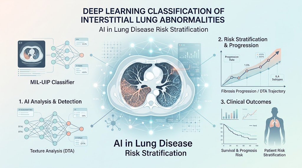
Deep learning classifier (MIL-UIP) applied to baseline CT scans objectively estimates usual interstitial pneumonia (UIP) likelihood in individuals with interstitial lung abnormalities (ILA).
MIL-UIP scores above 0.5 predict significant progression of fibrotic changes on follow-up CT, reflected by data-driven textural analysis (DTA).
Higher MIL-UIP scores associate independently with increased mortality across two large observational cohorts.
Automated imaging biomarkers hold promise for early risk stratification and enrichment of clinical trials targeting progressive interstitial lung disease.
Study Background
Interstitial lung abnormalities (ILA) are increasingly detected radiologically in populations undergoing chest computed tomography (CT) for various reasons. Although ILAs reflect subclinical interstitial lung disease (ILD) manifestations, their natural history is heterogeneous; some remain stable while others progress to fibrotic lung disease with significant morbidity and mortality. The clinical challenge lies in identifying individuals at high risk for progression and poor outcomes to guide monitoring and therapeutic intervention. Traditional visual CT scoring has limitations in reproducibility and scalability, driving interest in objective, automated imaging biomarkers. Patterns resembling usual interstitial pneumonia (UIP), the histopathological hallmark of idiopathic pulmonary fibrosis (IPF), confer a poor prognosis. Leveraging advances in artificial intelligence, this study investigates whether a deep learning classifier can quantify UIP-like radiologic features to prognosticate fibrosis progression and survival in individuals with ILA.
Study Design
This study analyzed baseline and serial follow-up chest CT scans from two large observational cohorts: COPDGene and AGES-Reykjavík, both comprising individuals without clinically diagnosed ILD at enrollment. The imaging datasets were processed using two complementary techniques: data-driven textural analysis (DTA) to objectively quantify fibrotic pattern extent and a deep learning-based multiple-instance learning UIP classifier (MIL-UIP) to estimate the likelihood of UIP-like features on baseline CT. The primary outcomes were longitudinal changes in fibrotic pattern by DTA and overall survival. Statistical associations between baseline MIL-UIP scores and these outcomes were assessed using linear mixed-effects models for fibrosis progression and multivariable Cox proportional hazards models for survival, adjusting for relevant covariates.
Key Findings
The deep learning classifier’s performance was consistent across cohorts. Baseline MIL-UIP scores exceeding 0.5 identified individuals with significant annual relative increases in fibrotic DTA measures: 13.41% (95% CI: 8.10%, 18.99%; p < 0.001) in COPDGene and 14.10% (95% CI: 5.06%, 23.91%; p = 0.002) in AGES-Reykjavík. Moreover, each 0.1 increment in MIL-UIP score correlated with a 1.10 percentage point (95% CI: 0.45, 1.74; p 0.5 conferred a significantly higher hazard of mortality: HR 1.61 (95% CI: 1.08, 2.38; p = 0.018) for COPDGene and HR 1.62 (95% CI: 1.01, 2.61; p = 0.046) for AGES-Reykjavík. These imaging biomarkers correlated closely with traditional visual assessments of ILA, UIP patterns, and fibrosis progression.
Expert Commentary
This study exemplifies the translational potential of artificial intelligence in thoracic imaging to augment clinical decision-making for ILD risk stratification. The MIL-UIP classifier offers a standardized, reproducible method to identify subclinical UIP-like features that predict fibrotic worsening and mortality in populations without overt ILD diagnoses. Its validation in two distinct cohorts supports generalizability. Notwithstanding, limitations include potential cohort-specific imaging acquisition variability and absence of histopathologic confirmation. Further prospective studies integrating clinical parameters and biomarker assays will elucidate how best to incorporate these automated tools into routine risk assessment and therapeutic trial enrichment.
Conclusion
Automated deep learning classification of UIP-like radiologic patterns on chest CT in individuals with ILA strongly associates with fibrosis progression and mortality. These findings endorse the utility of AI-based imaging biomarkers to objectively risk stratify patients at the earliest disease stages, potentially guiding surveillance intensity and enrollment criteria in clinical trials targeting progressive interstitial lung disease. Continued refinement and validation will be essential before clinical adoption.
Funding and ClinicalTrials.gov
The study was supported by research grants from the National Institutes of Health and other institutional sources associated with the COPDGene and AGES-Reykjavík cohorts. No clinical trial registration was reported as this analysis utilized observational cohort datasets.
References
1. Baraghoshi D, Thieke D, Hatabu H, et al. Deep learning classification of radiologic pattern is associated with progression of interstitial lung abnormalities and with survival. Am J Respir Crit Care Med. 2026;PMID: 42811099.
2. Hatabu H, Hunninghake GM, Richeldi L, et al. Interstitial lung abnormalities detected incidentally on CT: A pictorial review with main clinical and imaging features. Radiology. 2019;292(2):303-312.
3. Lederer DJ, Martinez FJ. Idiopathic pulmonary fibrosis. N Engl J Med. 2018;378(19):1811-1823.
4. Walsh SLF, Calandriello L, Sverzellati N, et al. Deep learning for classification of usual interstitial pneumonia: A validation study. Lancet Respir Med. 2020;8(11):1050-1059.