Highlight
- Uterine artery Doppler at 35-36 weeks reflects primarily placental angiogenic imbalance associated with preeclampsia.
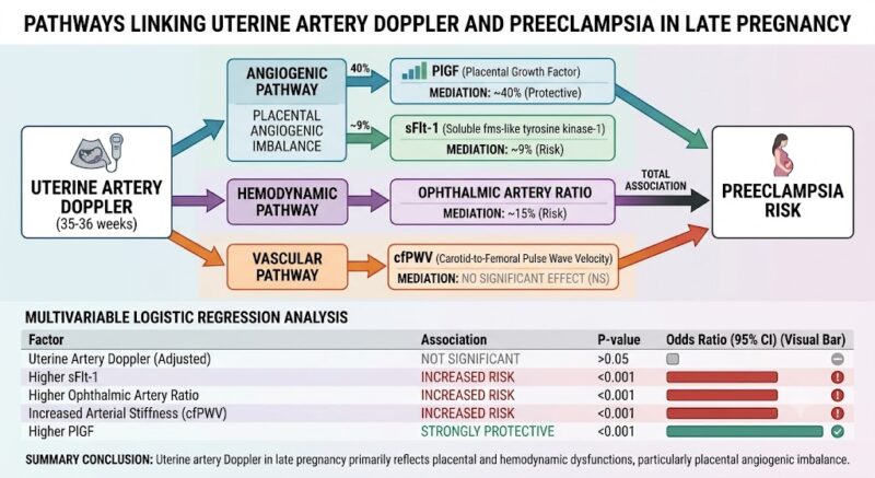
- Placental growth factor (PlGF) mediates approximately 40% of the association, with soluble fms-like tyrosine kinase-1 (sFlt-1) mediating about 9%.
- Ophthalmic artery hemodynamic changes account for 15% mediation, while arterial stiffness (cfPWV) shows no significant impact.
- After adjusting for angiogenic and vascular variables, the uterine artery Doppler pulsatility index loses independent predictive value for preeclampsia.

Study Background
Preeclampsia remains a major contributor to maternal and perinatal morbidity globally, characterized by new-onset hypertension and end-organ dysfunction during pregnancy. Its complex etiology involves abnormal placentation leading to impaired uteroplacental perfusion and systemic maternal vascular maladaptation. Early pregnancy uterine artery Doppler velocimetry is an established tool to screen for defective placentation, yet its role in late pregnancy assessment is less defined. Understanding what late pregnancy uterine artery Doppler indices signify biologically could help refine risk stratification and management strategies for preeclampsia.
Study Design
This prospective observational cohort study evaluated 11,962 singleton pregnancies at 35+0 to 36+6 weeks gestation in a tertiary maternity center in London from 2021 to 2024. Comprehensive assessments included:
- Uterine artery pulsatility index (PI) via Doppler ultrasound as a measure of placental vascular resistance.
- Quantification of placental angiogenic biomarkers: placental growth factor (PlGF) and soluble fms-like tyrosine kinase-1 (sFlt-1), key regulators implicated in preeclampsia pathogenesis.
- Hemodynamic evaluation via ophthalmic artery peak systolic velocity ratio, reflecting systemic vascular changes related to preeclampsia.
- Assessment of macrovascular function through carotid to femoral pulse wave velocity (cfPWV), an indicator of arterial stiffness.
The primary outcome was the occurrence of preeclampsia. Mediation analysis was employed to determine how much of the uterine artery Doppler’s association with preeclampsia was explained by angiogenic, hemodynamic, or vascular pathways. Multivariable logistic regression was used to control for confounders and evaluate independent predictors.
Key Findings
Preeclampsia was diagnosed in 349 pregnancies (2.9%). The main findings include:
1. Mediation by Placental Angiogenic Markers
Placental angiogenic imbalance had the largest mediating effect on the relationship between uterine artery Doppler and preeclampsia. Specifically:
- Decreased PlGF levels mediated approximately 40% of this association, confirming its central role in endothelial dysfunction and placental insufficiency.
- Elevated sFlt-1 mediated a smaller but significant 9% proportion, reflecting its antiangiogenic contribution.
2. Hemodynamic Influence
The ophthalmic artery peak systolic velocity ratio, a proxy for maternal systemic hemodynamic adaptation, mediated about 15% of the total association. This suggests that changes in maternal cerebral vascular resistance partially link uterine artery flow abnormalities with preeclampsia.
3. Arterial Stiffness
Carotid to femoral pulse wave velocity (cfPWV) did not significantly mediate the association between uterine artery Doppler findings and preeclampsia, indicating that large artery stiffness may not be a major pathway at this gestational stage.
4. Independent Associations
In multivariable logistic regression including uterine artery Doppler, angiogenic markers, hemodynamic indices, and arterial stiffness:
- Uterine artery PI was no longer independently predictive of preeclampsia (β=0.11, p=0.13), implying its effects are largely exerted through placental and hemodynamic pathways.
- Higher sFlt-1, elevated ophthalmic artery ratio, and increased arterial stiffness were independently associated with increased preeclampsia risk (all p<0.001).
- Higher PlGF held a strong protective association (p<0.001).
Expert Commentary
This comprehensive study leverages a large cohort and advanced causal mediation methodology to unpack the multifactorial nature of preeclampsia signals detected by uterine artery Doppler late in pregnancy. The predominant mediation by placental angiogenic imbalance substantiates the pivotal role of defective placentation beyond early gestation. The partial mediation by ophthalmic artery hemodynamics aligns with systemic vascular alterations in preeclampsia progression.
The lack of significant mediation by arterial stiffness may reflect the timing relative to disease onset or the primary microvascular rather than macrovascular involvement early in preeclampsia development. Clinicians should recognize that elevated uterine artery pulsatility late in pregnancy is a marker of complex placental and maternal cardiovascular dysfunction, rather than a stand-alone predictor.
Limitations include the observational design, potential residual confounding, and single-center setting that might affect generalizability. Confirmation in diverse populations and exploration of temporal dynamics of these markers with clinical outcomes will be valuable.
Conclusion
Late pregnancy uterine artery Doppler abnormalities in preeclampsia predominantly mirror placental angiogenic dysregulation and, to a lesser extent, maternal hemodynamic alterations. These findings refine the interpretation of Doppler results beyond early screening, suggesting potential integrative biomarker strategies for monitoring and managing high-risk pregnancies. Further studies to translate these mechanistic insights into tailored clinical interventions are warranted.
Funding and ClinicalTrials.gov Registration
Details on funding sources and trial registration were not provided in the published abstract. Further inquiry into the full article is advised for compliance and transparency details.
References
1. Chatzakis C, Mansukhani T, Bhojwani D, et al. What does uterine artery Doppler reflect in late pregnancy? Insights from angiogenic, hemodynamic, and vascular markers in preeclampsia. Am J Obstet Gynecol. 2026 Aug 7. PMID: 42567393.
2. Levine RJ, Maynard SE, Qian C, et al. Circulating angiogenic factors and the risk of preeclampsia. N Engl J Med. 2004;350(7):672-683.
3. Poon LC, Kametas NA, Chelemen T, et al. Maternal hemodynamics in normal pregnancy and preeclampsia assessed by Doppler ultrasound. Hypertension. 2009;54(1):94-100.
4. Vlachopoulos C, Aznaouridis K, Stefanadis C. Prediction of cardiovascular events and all-cause mortality with arterial stiffness: a systematic review and meta-analysis. J Am Coll Cardiol. 2010;55(13):1318-1327.