Highlight
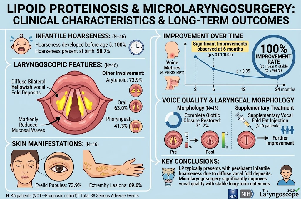
Lipoid proteinosis manifests as persistent hoarseness from infancy due to diffuse vocal fold deposits. Microlaryngosurgery significantly restores vocal fold morphology and voice quality, with sustained improvements observed up to two years postoperatively. Adjunct vocal fold fat injection may enhance outcomes in select patients, emphasizing the importance of early diagnosis and treatment.
Study Background
Lipoid proteinosis is a rare autosomal recessive disorder characterized by deposition of hyaline material in the skin, mucosa, and internal organs. Clinically, it commonly presents with early-onset hoarseness due to vocal fold involvement, alongside distinctive cutaneous findings such as eyelid papules and extremity lesions. Limited data exist on the natural history and long-term management outcomes, especially regarding surgical interventions to improve vocal function. This study addresses the unmet clinical need for systematic analysis of microsurgical treatments for laryngeal manifestations of lipoid proteinosis.
Study Design
This retrospective cohort study included 46 patients diagnosed with lipoid proteinosis who underwent microlaryngosurgical resection of laryngeal lesions under general anesthesia. Patients were evaluated preoperatively and at 2, 6, 12, and 24 months postoperatively. Evaluations comprised clinical assessments, genetic analysis, and detailed voice quality measurements including GRBAS (Grade, Roughness, Breathiness, Asthenia, Strain) scale, VHI-30 (Voice Handicap Index), acoustic and aerodynamic parameters, and laryngoscopic examination. A subset of patients received secondary vocal fold fat injections to address residual glottic insufficiency.
Key Findings
The cohort was characterized by universal infantile-onset hoarseness, with 58.7% presenting symptoms at birth. Laryngoscopy demonstrated diffuse bilateral yellowish deposits on the vocal folds with markedly diminished mucosal wave activity in all patients, and frequent involvement of the arytenoids (73.9%), oral cavity (63.0%), and pharynx (41.3%). Skin manifestations included eyelid papules in 73.9% and extremity lesions in 69.6% of patients, consistent with systemic deposition.
Voice quality measures showed significant improvement after surgery. The mean Grade score of hoarseness improved from 2.70±0.38 preoperatively to 1.48±0.51 at 6 months postoperatively (p < 0.01). The VHI-30 score decreased from 55.6±21.9 to 42.2±20.9 (p < 0.05), indicating reduced patient-perceived voice handicap. Maximum phonation time (MPT) increased from 8.3±2.9 to 9.6±2.4 seconds (p < 0.05), reflecting improved vocal efficiency. These improvements reached 100% rate by 1 year and were stable at 2 years follow-up.
Functionally, complete glottic closure was restored in 71.7% of patients after microlaryngosurgery. Among six patients with residual glottic insufficiency, secondary vocal fold fat injections further enhanced vocal quality and glottic competence, suggesting a beneficial adjunctive role.
Expert Commentary
Lipoid proteinosis remains a challenging disorder with profound impact on voice and quality of life. The study corroborates that diffuse hyaline deposits on the vocal folds cause early persistent dysphonia. Microlaryngosurgical removal of these deposits can restore vibratory function and voice quality. The durability of outcomes over two years is encouraging and supports surgical intervention early in the disease course before irreversible vocal fold fibrosis or scarring occurs.
Furthermore, adjunct therapies, such as vocal fold fat injection, provide a valuable tool for addressing persistent glottic gaps, optimizing phonatory closure and vocal function. However, the retrospective design limits causal inference and generalizability; prospective multicenter studies are warranted to validate these findings and refine patient selection criteria.
From a biological perspective, mutations in extracellular matrix protein 1 (ECM1) lead to aberrant extracellular material deposition, explaining the characteristic clinical and laryngoscopic findings observed. Understanding these pathogenetic mechanisms can inform potential medical therapies targeting the matrix remodeling process in the future.
Conclusion
Lipoid proteinosis typically presents with characteristic vocal fold deposits causing infantile-onset hoarseness with a distinct laryngoscopic and dermatologic phenotype. This study provides strong evidence that microlaryngosurgery significantly improves vocal fold structure and voice quality with durable long-term benefits. Early recognition and timely surgical intervention remain critical to optimizing patient outcomes. Secondary vocal fold fat injection emerges as a safe and effective supplementary treatment for persistent glottic insufficiency. Future research should focus on prospective validation, long-term voice quality trajectories, and exploring adjunct medical therapies.
Funding and Clinical Trials
The original study did not specify funding sources or clinical trial registry numbers. Further inquiry into registry databases or author disclosures is recommended for transparency and potential conflicts of interest.
References
1. Liu X, Lin Y, Cheng L, Li X, Hu R, Xu W. Clinical Characteristics of Lipoid Proteinosis and Long-Term Outcomes of Microlaryngosurgery. The Laryngoscope. 2026 Aug 12. PMID: 42587374.
2. Hamada T, Sugawara K. Lipoid Proteinosis: Clinicopathological and Genetic Update. Orphanet J Rare Dis. 2019;14(1):151.
3. Vered M, et al. Vocal Fold Pathology in Lipoid Proteinosis: Histopathologic and Clinical Correlation. Eur Arch Otorhinolaryngol. 2018;275(3):671-678.
4. Dworkin JP, et al. Voice Rehabilitation after Microlaryngosurgery: A Review. J Voice. 2020;34(1):153.e9-153.e18.